ISA KANYAKUMARI

ANAESTHESIA PEARLS


  SAFE ANAESTHESIA PRACTICE
  International Standards for a Safe Practice of Anaesthesia 2010
 
  This is the 2010 update of the Standards developed by the International Task Force on Anaesthesia Safety that were adopted by the World Federation of Societies of Anaesthesiologists 13 June 1992.
  These standards are recommended for anaesthesia professionals throughout the world.
  Standards that would be expected in all anaesthesia care for elective surgical procedures are termed "HIGHLY RECOMMENDED" and these are the functional equivalent of "mandatory" standards.
 
  These HIGHLY RECOMMENDED standards (regarding facilities, equipment, and medications) are necessary for "Level 1" or "basic" infrastructure to any healthcare environment anywhere in which general or regional anaesthetics are administered.
 
  In spite of some facilities' limitations, it may be possible to implement elements of the RECOMMENDED standards even in a "basic" setting and, likewise, to implement elements of the SUGGESTED standards even in an "intermediate" setting.
 
  Table for a detailed outline of the integration of the practice standards with the levels of facilities/infrastructure.
 
Anaesthesia standards (in order of adoption) SettingInfrastructure
HIGHLY RECOMMENDEDLevel 1Basic
HIGHLY RECOMMENDED + RECOMMENDEDLevel 2Intermediate
HIGHLY RECOMMENDED + RECOMMENDED + SuggestedLevel 3Optimal
 
HIGHLY RECOMMENDED RECOMMENDEDSUGGESTED
Oxygenation
Oxygen supply
Oxygenation of the patient :
Supplemental oxygen
Un interrupted supply
Visual examination
Adequate illumination
Pulse oximetry
Inspired oxygen concentration
Oxygen supply failure alarm
Hypoxic Guard
-
Airway and ventilation
 
Observation
Auscultation
The reservoir bag
Precordial
Pretracheal, or
Oesophageal stethoscope
Capnography
Continuous measurement of the inspiratory and/or expired gas volumes, and of the concentration of volatile agents
Circulation
Cardiac rate and rhythm
Tissue perfusion
Blood pressure
Palpation of the pulse
Auscultation of the heart sounds
Pulse oximetry
Clinical examination
At least every 5 mts
Electrocardiograph
Defibrillator
Capnography
NIBP
IABP
Temperature
 
At frequent intervals
Continual electronic temperature measurement
-
Neuromuscular function
 
-
Peripheral nerve stimulator
-
Depth of anaesthesia
 
Degree of unconsciousness (clinical observation)
Continuous measurement of the inspiratory and/or expired gas volumes, and of the concentration of volatile agents
BIS Monitor
Audible signals and alarms
 
Available audible signals (pulse tone of the pulse oximeter) and audible alarms (with appropriately set limit values) should be activated at all times and loud enough to be heard throughout the operating room
 -
-
  Table Guide to Infrastructure, Supplies and Anaesthesia Standards at Three Levels of Health Care Facility Infrastructure and Supplies.
  (Emergency and Essential Anaesthesia and Surgical Procedures, adapted in part from WHO Manual: Surgical Care at the District Hospital 2003 and the 1992 International Standards for a Safe Practice of Anaesthesia)
 
Level 1Level 2Level 3
(Should meet at least HIGHLY RECOMMENDED anaesthesia standards)(Should meet at least HIGHLY RECOMMENDED and RECOMMENDED anaesthesia standards)(Should meet at least HIGHLY RECOMMENDED, RECOMMENDED and SUGGESTED anaesthesia standards)
Small hospital / health centreDistrict/provincial hospitalReferral hospital
Rural hospital or health centre with a small number of beds (or urban location in an extremely disadvantaged area); sparsely equipped operating room (OR) for "minor" procedures
Provides emergency measures in the treatment of 90-95% of trauma and obstetrics cases (excluding caesarean section)
Referral of other patients (for example, obstructed labour, bowel obstruction) for further management at a higher level
District or provincial hospital (e.g. with100-300 beds) and adequately equipped major and minor operating rooms
Short term treatment of 95-99% of the major life threatening conditions
A referral hospital of 300-1000 or more beds with basic intensive care facilities. Treatment aims are the same as for Level 2, with the addition of:
Ventilation in OR and ICU
Prolonged endotracheal intubation
Thoracic trauma care
Haemodynamic and inotropic treatment
Basic ICU patient management and monitoring for up to 1 week : all types of cases, but possibly with limited provision for:
Multi-organ system failure
Haemodialysis
Complex neurological and cardiac surgery
Prolonged respiratory failure
Metabolic care or monitoring
Essential ProceduresEssential ProceduresEssential Procedures
Normal delivery
Uterine evacuation
Circumcision
Hydrocele reduction, incision and drainage
Wound suturing
Control of haemorrhage with pressure dressings
Debridement and dressing of wounds
Temporary reduction of fractures
Cleaning or stabilization of open and closed fractures
Chest drainage (possibly)
Abscess drainage
Same as Level 1 with the following additions:
Caesarean section
Laparotomy (usually not for bowel obstruction)
Amputation
Hernia repair
Tubal ligation
Closed fracture treatment and application of plaster of Paris
Acute open orthopaedic surgery: e.g internal fixation of fractures
Eye operations, including cataract extraction
Removal of foreign bodies: e.g. in the airway
Emergency ventilation and airway management for referred patients such as those with chest and head injuries
Same as Level 2 with the following additions:
Facial and intracranial surgery
Bowel surgery
Paediatric and neonatal surgery
Thoracic surgery
Major eye surgery
Major gynaecological surgery, e.g. vesico-vaginal repair
PersonnelPersonnelPersonnel
Paramedical staff/anaesthetic officer (including on-the-job training) who may have other duties as well
Nurse-midwife
One or more trained anaesthesia professionals
District medical officers, senior clinical officers, nurses, midwives
Visiting specialists or resident surgeon and/or obstetrician/ gynaecologist
Clinical officers and specialists in anaesthesia and surgery
DrugsDrugsDrugs
Ketamine 50 mg/ml injection
Lidocaine 1% or 2%
Diazepam 5 mg/ml injection, 2 ml or midazolam 1mg/ml injection, 5 ml
Pethidine 50 mg/ml injection, 2 ml
Morphine 10mg/ml, 1 ml
Epinephrine (Adrenaline) 1 mg
Atropine 0.6 mg/ml
Appropriate inhalation anaesthetic if vaporizer available
Same as Level 1 with the following additions:
Thiopental 500 mg/1g powder or propofol.
Suxamethonium bromide 500 mg powder
Pancuronium
Neostigmine 2.5 mg injection
Ether, halothane or other inhalation anaesthetics
Lidocaine 5% heavy spinal solution, 2 ml
Bupivacaine 0.5% heavy or plain, 4 ml
Hydralazine 20 mg injection
Frusemide 20 mg injection
Dextrose 50% 20 ml injection
Aminophylline 250 mg injection
Ephedrine 30/50 mg ampoules
Hydrocortisone
(?) Nitrous oxide
Same as Level 2 with the following additions:
Propofol
Nitrous oxide
Various modern neuromuscular blocking agents
Various modern inhalation anaesthetics
Various inotropic angents
Various intravenous antiarrhythmic agents
Nitroglycerine for infusion
Calcium chloride 10% 10 im injection
Potassium chloride 20% 10 ml injection for infusion
Equipment: capital outlayEquipment: capital outlayEquipment: capital outlay
Adult and paediatric self-inflating breathing bags with masks
Foot-powered suction
Stethoscope, sphygmomanometer, thermometer
Pulse oximeter
Oxygen concentrator or tank oxygen and a draw-over vaporizer with hoses
Laryngoscopes, bougies
Complete anaesthesia, resuscitation and airway management systems including:
Reliable oxygen sources
Vaporizer(s)
Hoses and valve
Bellows or bag to inflate lungs
Face masks (sizes 00-5)
Work surface and storage
Paediatric anaesthesia system
Oxygen supply failure alarm; oxygen analyzer
Adult and paediatric resuscitator sets
Pulse oximeter, spare probes, adult and paediatric*
Capnograph*
Defibrillator (one per O.R. suite / ICU)*
ECG (electrocardiograph) monitor*
Laryngoscope, Macintosh blades 1-3(4)
Oxygen concentrator[s] [cylinder]
Foot or electric suction
IV pressure infusor bag
Adult and paediatric resuscitator sets
Magill forceps (adult and child), intubation stylet and/or bougie
Spinal needles 25G
Nerve stimulator
Automatic non-invasive blood pressure monitor
Same as Level 2 with these additions (per each per OR room or per ICU bed, except where stated):
ECG (electrocardiograph) monitor*
Anaesthesia ventilator, reliable electric power source with manual override
Infusion pumps (2 per bed)
Pressure bag for IV infusion
Electric or pneumatic suction
Oxygen analyzer*
Thermometer [temperature probe*]
Electric warming blanket
Electric overhead heater
Infant incubator
Laryngeal mask airways sizes 2, 3, 4 (3 sets per O.R)
Intubating bougies, adult and child (1 set per O.R)
Anaesthetic agent (gas and vapour) analyser
Depth of anaesthesia monitor are being increasingly recommended for cases at high risk of awareness but are not standard monitoring in many countries.
Equipment: disposableEquipment: disposableEquipment: disposable
Examination gloves
IV infusion/drug injection equipment
Suction catheters size 16 FG
Airway support equipment, including airways and tracheal tubes
Oral and nasal airways
ECG electrodes
IV equipment (minimum fluids: normal saline, Ringer's lactate and dextrose 5%)
Paediatric giving sets
Suction catheters size 16 FG
Sterile gloves sizes 6-8
Nasogastric tubes sizes 10-16 FG
Oral airways sizes 000-4
Tracheal tubes sizes 3-8.5 mm
Spinal needles sizes 22 G and 25G
Batteries size C
Same as Level 2 with these additions:
Ventilator circuits
Yankauer suckers
Giving sets for IV infusion pumps
Disposables for suction machines
Disposables for capnography, oxygen analyzer, in accordance with manufacturers' specifications:
Sampling lines
Water traps
Connectors
Filters - Fuel cells
  Ref:
  International Standards for a Safe Practice of Anesthesia 2010. Canadian Journal of Anesthesia: November 2010, Volume 57, Issue 11, pp 1027-1034
  Advanced hemodynamic monitoring
  Introduction
  Hemodynamic monitoring is a cornerstone of care for the hemodynamically unstable patient.It is routinely used in the operating room during high-risk surgery.
  Hemodynamic monitoring comes from an understanding of the pathophysiology of the process being treated, such as heart failure or hypovolemic shock.
  New Concepts in Hemodynamic Monitoring
  Circulatory shock is defined by decreased ability of blood flow to meet the metabolic demands of the body. Four basic groups of circulatory shock can be defined: Hypovolemic, Cardiogenic, Obstructive, and Distributive.
  Tissue hypo-perfusion is common in all forms of shock (with the possible exception of hyperdynamic septic shock). Because specific types of circulatory shock require different therapies and target end-points of resuscitation, defining the cardiovascular state is important in determining both treatment options and their goals.
  Much of the rationale for hemodynamic monitoring resides at this level.
 
  Hemodynamic Profiles in Shock
  Certain combinations of hemodynamic findings allow the etiology of circulatory shock to be defined using this nosology.
 
Class of Shock CVP PAOP CO/CI SVR
Cardiogenic CVP PAOP CO/CI SVR
Hypovolemic CVP PAOP CO/CI SVR
Hyperdynamic septic CVP PAOP CO/CI SVR
Hypodynamic septic CVP PAOP CO/CI SVR
  The ultimate goal of management of shock is to maintain “Tissue Perfusion” which is monitored by “Adequate Oxygen Delivery”.
  Oxygen demand > Oxygen delivery
  Review of Basics:
  Oxygen Delivery (DaO2) = CaO2 x CO x 10 = 1000 ml/min
  Arterial Oxygen Content (CaO2) = (0.0138 x Hgb x SaO2) + (0.0031 x PaO2) = 20.1 ml/dl
  Cardiac Output (CO) = SV x HR =4-8 L/min
  Mixed venous (SvO2) and central venous oxygen saturations (ScvO2) reflect the balance between oxygen requirement and oxygen delivery, and thus may be used to assess the adequacy of tissue oxygenation.
  Venous oximetry allows the critical estimation of the global oxygen (O2) supply-demand ratio and can be gained from mixed (SvO2) and central venous blood (ScvO2).
 
SvO2 - True mixed venous oxygen saturation (60-80%)
ScvO2 - Central venous oxygen saturation (70%)
VO2 – Consumption of oxygen (200-250ml O2/min)
DO2 – Delivery of oxygen (950- 1150 ml O2/min)
  Pulmonary artery catheterization allows obtaining true mixed venous oxygen saturation
  (SvO2) while measuring central venous oxygen saturation (ScvO2) via central venous catheter reflects principally the degree of oxygen extraction from the brain and the upper part of the body.
  (For details of Venous Oximetry refer Anaesthesia Pearls Feb 2013- http://www.isakanyakumari.com/february-1-2013.php)
 
  Continuous SvO2/ScvO2 Monitoring:
 
  A decrease in SvO2 and ScvO2 represents an increased metabolic stress, either DO2 does not increase in such a way to cover an increased VO2, or DO2 drops because of decrease in either arterial O2 content, cardiac output, or both. The magnitude of the decrease indicates the extent to which the physiological reserves are stressed.
  How do we measure SvO2% & ScvO2%?
  Swan Ganz Pulmonary Artery Catheter (Old method)
  Advanced Technology Catheters:
 
CCO (Continuous CO)
CCOmbo (Continuous CO + Venous Oximetry)
CCOmbo/VIP
CCOmbo/EDV
SvO2
CCO/CEDV
CCOmbo/CEDV
CCOmbo/CEDV/VIP
 
Parameters Derived Information
SvO2 (Mixed venous Oxygen Saturation) Tissue Oxygenation
CEDV (Continuous End Diastolic Volume) Preload
SVR (Systemic Vascular Resistance) Afterload
CCO (Continuous Cardiac Output) Contractility
SV (Stroke Volume) Contractility
REVF (Right Ventricular Ejection Fraction) Contractility
  Normal Hemodynamic Values
 
SVO2 60-75%
Stroke volume 50-100 mL
Stroke index 25-45 mL/M2
Cardiac output 4-8 L/min
Cardiac index 2.5-4.0 L/min/M2
MAP 60-100 mm Hg
CVP 2-6 mm Hg
PAP systolic 20-30 mm Hg
PAP diastolic 5-15 mm Hg
PAOP (wedge) 8-12 mm Hg
SVR 900-1300 dynes.sec.cm-5
  These parameters can be used for Early Goal-Directed Therapy Treatment Protocol:
 
  How to assess volume responsiveness?
 
Stroke Volume Variations (SVV)
Pulse Pressure Variations (PPV)
Systolic Pressure Variations (SPV)
  Volume Responsiveness was defined as increase in CO by 15% or more, by 500 ml fluid bolus or by Passive Leg Raising.
  The volume responsiveness can also assessed by CVP-MAP Relationship or CVP vs. EDV/EDVI.
  New Generation Monitors uses Stroke Volume Variations, Pulse Pressure Variations and Systolic Pressure Variations for assessment of volume responsiveness.
  Stroke Volume Variation - FloTrac Vigileo:
  (For details of SVV refer Anaesthesia Pearls Feb 2013-http://www.isakanyakumari.com/february-1-2013.php)
 
  SVV can be used as a tool for volume responsiveness in low CO states.
  SVV > 13% = Volume Responsive.
  “SVV and PPV are more effective indicators for Volume Responsiveness than static indicators of preload (CVP, PAOP).”
  Limitations:
 
Patient needs to be on 100% Controlled Mechanical Ventilation.
Spontaneous Ventilation
Arrhythmias can affect SVV.
  Hemodynamic Monitoring
  Static Hemodynamic Monitoring:
 
Heart Rate
Blood Pressure ( Noninvasive vs. Invasive)
Central Venous Pressure.
The pulmonary artery catheter (PAC)
  Functional hemodynamic monitoring:
 
Volume challenge
Passive leg raising
Changes in central venous pressure during spontaneous breathing
Changes in left ventricular output during positive pressure ventilation
  Advanced hemodynamic monitoring:
 
Cardiac Output Measurement – CCO, CCI, SV, SVI, SVV
Tissue Oxygen Saturation Measurements- SvO2 or ScvO2
Echocardiography (TTE, TEE.)
Thoracic Bioimpedance
Thoracic Bioreactance
Endotracheal CO Monitor
The NICO System (Fick’s Principle for CO2)
Esophageal Doppler
 
  Clinical caveats for hemodynamic variables
 
Type of hemodynamic variable Parameter Comments
Solitary Blood pressure Hypotension is always pathological
  Central venous pressure (CVP) CVP is only elevated in disease
  Pulmonary artery occlusion pressure (Ppao) Ppao is the back-pressure to pulmonary blood flow
  Cardiac output There is no normal cardiac output, only an adequate or inadequate one
  Mixed venous oxygen saturation (SvO2) Decreasing SvO2 is a sensitive but nonspecific marker of cardiovascular stress
Dynamic Volume challenge Positive response defined as an increase in any of blood pressure, CVP, Ppao, cardiac output and/or SvO2, or a decrease in heart rate
  Echocardiographic analysis of vena cavae collapse during positive pressure inspiration identifies CVP <10 mmHg if it detects Complete inferior vena caval collapse a
>36% collapse in superior vena cavaa
  Defining preload responsiveness ≥13% pulse pressure variation during positive pressure ventilationa
>1 mmHg decrease in CVP during spontaneous inspirationb
a - Requires a fixed tidal volume of 6–8 ml/kg and complete adaptation to the ventilator
b- Requires a spontaneous inspiratory effort greater than –2 mmHg to be valid.
(Source: Critical Care December 2005 Vol 9 No 6 Pinsky and Payen)
  Central venous pressure (CVP)
  CVP can reflect a volume increase in RA pressures or decrease in RV contractility, can be both. Need to be monitored in conjunction with other monitors. (CVP&MAP)
  The main limitations of CVP monitoring:
 
It does not allow to measure cardiac output
It does not provide reliable information on the status of the pulmonary circulation in the presence of left ventricular dysfunction.
  Measurement of Cardiac Output:
  Thermo Dilution (TD)
 
Dilution of Temperature (Cold NS)
Area Under the Curve (AUC)
Pulmonary Artery Catheter ( PACs) or
Newer Generation CVL w sensors
Intermittent vs. Continuous TD
  Dye/Indicator Dilution
 
Same Technique ( Dilution of dye or indicator instead of NS)
Dye(indocyanine green) vs. Indicator( Lithium)
A line vs. CVL , no need for PACs.
  Arterial Pulse Pressure Analysis
  Pulmonary Artery Catheters (PACs):
 
Both Thermo Dilution and Dye/Indicator Dilution require PACs to calculate Cardiac output.
The KEY advantage is simultaneous measurements of other hemodynamic parameters:
Cardiac Output(CO)
Pulmonary Artery Pressures ( PAD, PAOP)
Left Sided Filling
SvO2%
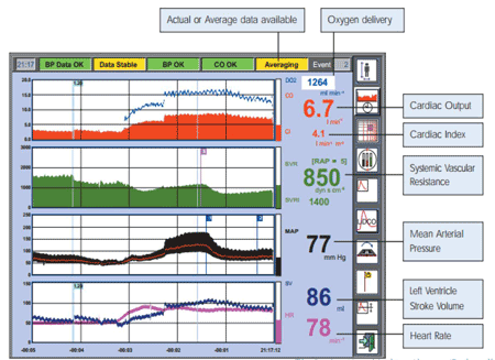
  The highlighted parameters provided by the Swan-Ganz Adva nced Technology pulmonary artery catheter DELIVER the most comprehensive view of oxygen flow and consumption:
 
  Source: Swan-Ganz Brochure, Edwards Critical Care Education
  Other ways of measuring CO by dilution technique:
  Transpulmonary Thermodilution Methods
 
PiCCO & PiCCO2 (Pulsion Medical Systems)
VolumeView (Edwards Life Sciences)
Intermittent vs. Continuous TD
  Lithium Dilution Technique
 
LiDCO /LiDCOplus/LiDCOrapid ( LiDCO limited)
  Ultrasound Indicator Dilution
 
COstatus (Transonic Systems, Inc.)
  COstatus (Transonic Systems, Inc.)
  With a single bolus of saline, provides key hemodynamic parameters including the following:
  Cardiac Function
 
Cardiac Output (CO)
Cardiac Index (CI)
Stroke Volume Index (SVI)
Total Ejection Fraction (TEF)
Systemic Vascular Resistance Index (SVRI)
  Blood Volumes
 
Total End Diastolic Volume Index (TEDVI)
Central Blood Volume Index (CBVI)
Active Circulation Volume Index (ACVI)
  Shunt Detection
 
Identifies shunts with direction of flow
 
  Source: http://www.transonic.com/ICU/COstatusSetup
  Less Invasive Methods for CO Calculation
  They are Arterial Waveform Analysis - Waveform Derived CO Measurements.
 
“BP is a product of SV (CO) and Vascular Resistance.”
Advantages:
Non invasiveness
Works through an already existing aline catheter
Continuous CO monitoring
May require calibration for arterial compliance and resistance:
Lithium Dilution ( LiDCO)
Thermodilution (VolumeView, PiCCO)
 
  Post–Cardiac Arrest Care
  2010 American Heart Association Guidelines for Cardiopulmonary Resuscitation and Emergency Cardiovascular Care.
  Introduction
 
Increasing recognition of return of spontaneous circulation (ROSC) - good quality of life post cardiac arrest.
Post–cardiac arrest care has significant potential to reduce early mortality caused by hemodynamic instability and later morbidity and mortality from multiorgan failure and brain injury.
Post–cardiac arrest care is a critical component of advanced life support.
Most deaths occur during the first 24 hours after cardiac arrest.
Multiple organ systems are affected after cardiac arrest.
  Three Critical Interventions to save lives in Cardiac Arrest:
 
Bystander CPR.
  Chest compressions only
Bystander CPR.
  “Cardio-cerebral resuscitation”
  Ventilation kills
Modern post-resuscitation care
  Therapeutic hypothermia
  Cardiac and hemodynamic support
  Cause of Death in Outside Hospital Cardiac Arrest (OHCA) and Inside Hospital Cardiac Arrest (IHCA)
 
  Mechanisms of brain injury in circulatory arrest
 
Primary Injury:
  “Energy failure” due to ATP depletion.
Secondary injury (6-72 Hrs)
  Loss of trans cellular electrolyte gradients.
    Ca+, Na+, Cl- enter, K+ exits cell.
    Water follows Na+ into cells causing cytotoxic edema.
  Lipid peroxidases damage membranes.
  Neurotransmitter release causes excitotoxicity.
  Activation of apoptotic pathways.
  Microvascular thrombosis.
  Reperfusion injury.
  Uncontrolled seizure activity.
  Hypotension, hypoperfusion.
  Post-resuscitation syndrome.
  ICP crisis.
  Auto regulatory failure.
  Fever.
  Re-arrest.
  Hypoxia.
  Derangements of glucose metabolism.
  Cardiac arrest associated brain injury “CAABI”
 
No flow” affects the most metabolically active areas of brain.
  Cortex.
  Basal ganglia.
  Cerebellum.
“Low flow” affects the watershed areas between vascular territories.
  Post-Cardiac Arrest Syndrome
  Post resuscitation disease is characterised by high levels of circulating cytokines and adhesion molecules, the presence of plasma endotoxins and deregulated leukocyte production of cytokines: a profile similar to that seen in severe sepsis.
 
Ischemia-refusion injury.
Systemic inflammatory response.
Multiorgan dysfunction.
May last for many days.
Organ support influences prognosis.
  Interventions
  Goal-Directed Strategies for Improving Outcome.
 
°C   Hypothermia.
O2    Normal Oxygenation.
CO2    Normal Ventilation.
BP    Hemodynamic Optimization.
ST    Coronary Reperfusion.
GL    Moderate Glycemic Control.
  Objectives of post–cardiac arrest care
 
Control body temperature to optimize survival and neurological recovery.
Identify and treat acute coronary syndromes (ACS).
Optimize mechanical ventilation to minimize lung injury.
Reduce the risk of multiorgan injury and support organ function if required.
Objectively assess prognosis for recovery.
Assist survivors with rehabilitation services when required.
Avoid using ties that pass circumferentially around the patient's neck.
Elevate the head of the bed 30°.
Avoid 100% oxygen - titrate inspired oxygen SpO2 of >94%, oxygen toxicity.
Avoid Hyperventilation or "overbagging" the patient.
 
Assess vital signs and monitor for recurrent cardiac arrhythmias.
Intravenous / intraosseous access.
Systolic blood pressure < 90 mm Hg- fluid boluses can be considered.
Titrated vasoactive drug infusions - minimum SBP (90 mm Hg) or a MAP (65 mm Hg).
Therapeutic hypothermia.
Coronary reperfusion (PCI).
 
  Hypothermia
  Clinical criteria for therapeutic hypothermia:
 
  No more than 8 hours have elapsed since the return of spontaneous circulation.
  Encephalopathy is present, typically defined as the patient being unable to follow verbal commands.
  There is no life-threatening infection or bleeding.
  Aggressive care is warranted and desired by the patient or the patient’s surrogate decision-maker.
Cooling delays cell death
Reduces cerebral metabolism.
Reduces inflammatory response.
Cooling improved neurological outcome after cardiac arrest.
32-34 degrees for 12-24 hours .
  Therapeutic Hypothermia
 
Cooling phases
Induction.
Maintenance.
Rewarming.
Methods
Internal – cold saline infusion, CPB, intravascular.
Heat exchangers.
External – ice packs, cooling blankets, water.
Circulating blankets and gel pads.
Cooling facilitated by sedation, paralysis, Mg
  Cooling Methods
 
  External Cooling
 
  Potential complications
 
Coagulopathy.
Arrhythmias.
Hyperglycemia.
Pneumonia .
Sepsis.
Infections.
Decrease immune function.
Impairs coagulation .
Ongoing bleeding.
  Oxygenation/ Normal Ventilation
 
  Titrate oxygen administration - maintain the SpO2 94%.
  Controlled ventilation with specific goals to keep PaCO2 37.6 to 45.1 mm Hg (5 to 6 kPa) and SaO2 95% to 98%.
  TV of 6 to 8 mL/kg and inspiratory plateau pressure 30 cm H2O to reduce ventilator-associated lung injury.
  Hemodynamic Optimization
 
Hemodynamic instability is common after cardiac arrest.
Mean arterial pressure 65 mm Hg,
CVP >8 < 20 mmHg and
ScvO2 70% are generally considered reasonable goals.
  Coronary Reperfusion
  Acute Coronary Syndrome (ACS) is a common cause of cardiac arrest diagnosed by 12-lead ECG and cardiac markers. Aggressive treatment of STEMI - regardless of coma or induced hypothermia with emergency coronary angiography is a must.
  PCI alone or as part of a bundle of care - improves myocardial function and neurological outcomes.
  Therapeutic hypothermia is safely combined with primary PCI after cardiac arrest caused by AMI.
  Antiarrhythmic drugs lidocaine or amiodarone are usually used.
  Moderate Glycemic Control
 
The post–cardiac arrest patient is likely to develop metabolic abnormalities such as hyperglycemia.
Increased mortality or worse neurological outcomes.
Optimum blood glucose concentration and interventional strategy to manage blood glucose in the post–cardiac arrest period is unknown.
Hypoglycemia may be associated with worse outcomes in critically ill patients.
Strategies to target moderate glycemic control (144 to 180 mg/dL) may be considered in adult patients with ROSC after cardiac arrest.
  Attempts to control glucose concentration within a lower range (80 to 110 mg/dL should not be implemented after cardiac arrest due to the increased risk of hypoglycaemia.
  Post‐Cardiac Arrest Syndrome - Goal‐Directed Strategies
 
  Goal For Improving Outcomes
°C Core Temp 32-34 °C ≤4 hr (or pre-PCI)
O2 Pulse Ox 94-98% ≤10 min
CO2 PetCO235-40 mmHg ≤20 min
BP MAP > 65 mm Hg ≤60 min
ST Coronary Reperfusion ≤90 min
GL Glucose < 180 mg/dl ?
  Steroids
 
Corticosteroids have an essential role in the physiological response to severe stress, including maintenance of vascular tone and capillary permeability.
In the post–cardiac arrest phase - adrenal insufficiency.
At present there are no human randomized trials investigating corticosteroid use after ROSC.
  Summary
  The goal of immediate post–cardiac arrest care is to optimize systemic perfusion, restore metabolic homeostasis, and support organ system function to increase the likelihood of intact neurological survival.
 

Ref:

  1. Mild therapeutic hypothermia to improve the neurologic outcome after cardiac arrest.N Engl J Med. 2002 Feb 21;346(8):549-56.
  2. Treatment of comatose survivors of out-of-hospital cardiac arrest with induced hypothermia.N Engl J Med. 2002 Feb 21;346(8):557-63.
  3. Mode of death after admission to an intensive care unit following cardiac arrest. November 2004, Volume 30, Issue 11, pp 2126-2128.
  4. Neurologic Prognosis in Cardiac Arrest Patients Treated With Therapeutic Hypothermia The Neurologist, Volume 17, Number 5, September.
  5. The use Hypothermia after cardiac arrest. Volume 38-Nov-Dec 1959. Anesthesia and Analgesia 1959;38 (6): 423
  6. Mild therapeutic hypothermia to improve neurological outcome after cardiac arrest. N Engl J Med 2002;346:549-56.
  7. Treatment of comatose survivors of out-of-hospital cardiac arrest with induced hypothermia. New Engl J Med 2002; 346:557-63.
  8. Therapeutic hypothermia: is it effective for non-VF/VT cardiac arrest? Critical Care 2013, 17:215.
  9. Outcome, timing and adverse events in therapeutic hypothermia after out-of-hospital cardiac arrest. Acta Anaesthesiologica Scandinavica, Volume 53, Issue 7, pages 926–934, August 2009.
  10. Post Resuscitation Care: Dr Loo Chian Min, Department of Respiratory & Critical Care Medicine Singapore General Hospital.
  11. Post‐Cardiac Arrest Syndrome Goal‐Directed Strategies for Improving Outcomes: Robert W. Neumar, MD, PhD, FACEP, Associate Professor of Emergency Medicine, Associate Director, Center for Resuscitation Science, University of Pennsylvania School of Medicine.
  12. Post-resuscitation care of the cardiac arrest survivor. David Seder MD., Maine Medical Center, Director of Neurocritical Care.
  13. 2010 American Heart Association Guidelines for Cardiopulmonary Resuscitation and Emergency Cardiovascular Care Science. Part 9: Post–Cardiac Arrest Care.
  PiCCO
  System Requires:
  Thermistor Tipped Aline Catheter –Femoral Aline ( longer cath, better signal), Radial, Axillary, Brachial as well. Central Venous Catheter (thermo indicator solution injection).
  The PiCCO-Technology parameters give answers to the following questions:
  1. What is the cardiovascular status?
 Cardiac Output (CO) - The Cardiac Output (CO) of the transpulmonary thermodilution is averaged over several respiration cycles while the bolus saline passes from the CVC to the arterial access.
  2. What is the cardiac preload?
  Global End-Diastolic Volume (GEDV) - represents the total of the 4 individual chambers of the heart and therefore reflects the cardiac preload. (The thorax has only a limited ability to expand and three intra-thoracic compartments interact with each other: filling of the ventricles (Global End-Diastolic Volume, GEDV), the Extravascular Lung Water (EVLW), and the gas volume (tidal volume/PEEP). If one of these components changes under otherwise stable conditions it will inevitably influence the others.)
  3. Will volume loading increase cardiac output?
  Stroke Volume Variation (SVV) gives – provided there is a fully ventilated patient with a stable heart rhythm – information as to whether an increase in preload will lead to an increased cardiac output.
  Change in preload leads to a variation in stroke volume in the case of a volume responsive heart.
 
  The difference in preload between inspiration and expiration caused by mechanical ventilation leads to different stroke volumes. This depends on which part of the Frank-Starling curve the function of the heart is situated.
 
  The increase in preload is identical: ΔEDV1 = ΔEDV2, but the influence of the stroke volume is different: ΔSV1 >> ΔSV2
  4. What is the afterload?
  Systemic Vascular Resistance (SVR) - The PiCCO-Technology provides the possibility of monitoring and managing the interaction between cardiac output and vascular resistance by continuously calculating both the cardiac output and the Systemic Vascular Resistance (SVR).
  5. What is the cardiac contractility?
  Pressure Velocity Increase (dPmx)
  Global Ejection Fraction (GEF)
  Optimization of fluid balance alone is not sufficient to stabilize the hemodynamic situation and to ensure adequate organ perfusion. Cardiac contractility and afterload are also important determinants of the Frank-Starling mechanism. The possible cardiac effects caused by different therapies are highlighted by the Global Ejection Fraction (GEF) and by measurement of the Left Ventricular Contractility (dPmx).
  6. Is lung edema developing or already present?
  Extravascular Lung Water (EVLW) - describes the amount of liquid in the lung tissue and marks the point where further volume loading is no longer an advantage, or requires critical balancing. An additional parameter of the PiCCO-Technology, the Pulmonary Vascular Permeability Index (PVPI), determines the cause of pulmonary edema. A high permeability index indicates capillary leakage due to an inflammatory process. If this index is normal, it is very likely a congestive-caused or cardiac-caused edema.
  EVLW and PVPI are of great significance in the treatment of cardiac or septic shock.
 
 
  LiDCOplus
  Combination of two monitors:
 
LiDCO (Indicator dilution CO calculation & monitor)
PulseCO (Software, CO from aline waveform.)
  This unique combination provides beat-to-beat measurement of cardiac output with lower risk and high precision.
  The PulseCO software calculates the pulse power and derived stroke volume from the arterial waveform. This avoids the necessity for detection of any particular waveform features such as the dichrotic notch. Thereby damping effect is also minimized in LiDCO system.
  Requires:
 
Arterial line (aline)
Peripheral or Central Access (to insert a lithium sensitive sensor)
  External CO calibration with lithium every 8 hours is necessary.
  LiDCOplus Screen shots:
 
 
  LiDCOrapid
  “No need for CO calibration with lithium. Replaced by nomogram which is derived form in vivo data to estimate CO”
  The nominal SV and CO are established from the arterial pressure waveform which is taken via simple cable connection from the vital signs monitor. Using the PulseCO algorithm the pressure is then converted into nominal SV and HR to a give CO that is then scaled to the patient's own characteristics.
  The LiDCOrapid displays the following parameters:
 
Pressures – MAP, Systolic and Diastolic
Heart Rate
Stroke Volume and Cardiac Output (Scaled or Actual)
Dynamic Preload parameters – Pulse Pressure Variation (PPV) and Stroke Volume Variation (SVV)
User selected event response window
 
  LiDCOrapidv2 with Unity Software
  LiDCO added the display of both continuous non invasive blood pressure (CNAP) and level of consciousness (BIS) to the LiDCOrapidv2 monitor platform.
 
 
FloTrac Vigileo
With the 3rd generation FloTrac system from Edwards Lifesciences, you’ll have access to automatic, up-to-the-minute cardiac output, stroke volume, stroke volume variation and systemic vascular resistance–under more patient conditions. The FloTrac sensor easily connects to any existing arterial catheter, and requires no manual calibration, making it the easy and reliable solution for fluid management. (Provides CO/CI, SV/SVI, SVV and SVR/SVRI)
  This latest enhancement evolves the algorithm using an expanded patient database. This database informs the algorithm to recognize and adjust for more patient conditions – including hyperdynamic conditions.
 
 
  Advantages:
 
Requires No Manual Calibration for CO Calculation
User enters Patient (Pt) Specific Data. ( age, gender, height, weight to initiate the monitoring.)
Advanced Arterial Waveform Analysis ( PRAM) by FloTrac Sensor
Pt to pt differences in vasculature
Real time changes in vascular tone
Different arterial sites are acceptable
Central Venous Oximetry (Scvo2) is available
  Disadvantages:
 
Aline Wave form is important !
Good Arterial Signal Quality is critical for accurate CO calculation.
Still Not Reliable
During Arrhythmias
For Hemodynamically Unstable Patients
Intra Aortic Balloon Pump in use.
Ventricular Assist Devices in use
  EV1000 Clinical Platform
  The EV1000 clinical platform presents a holistic view of clinically validated parameters provided by the FloTrac sensor, the PreSep and PediaSat oximetry catheters. With the EV1000 clinical platform, you are able to select parameters and the associated level of invasiveness.
  Color-based indicators communicate patient status at a glance, and visual clinical support screens allow for immediate recognition and increased understanding of rapidly changing clinical situations for improved decision enablement.
  Screen shots:
 
 
  Conclusion:
 
  The implementation of this monitor will allow for hemodynamic monitoring in patients who may not have previously been considered candidates due to risks versus benefits.
  The key number to treat for SVV is >13%. This value will provide information assisting the anesthesia provider if the patient needs intravascular volume, pressor agents, inotropes, or diuretics, based on the volume responsive algorithm.
  CO values from the Vigileo /FloTrac system trend in a similar manner to PAC. CO can be used confidently for trending hemodynamic data.
  Clinical judgment when utilizing this monitoring system is of utmost importance. The entire clinical picture must be analyzed before deciding on interventions that the patient requires.
  Optimization of hemodynamic variables with the assistance of the Vigileo/FloTrac monitoring system may improve patient outcomes.
  Hemodynamic monitoring of CO, CI, and SVV may now be available to a larger patient population with the utilization of the minimally invasive monitoring system.
  Ref:
  1. Update on Hemodynamic Monitoring. Gozde Demiralp, MD., Assistant Professor of Anesthesiology, Department of Anesthesiology, Division of Critical Care Medicine, University of Oklahoma, College of Medicine, Oklahoma City, Oklahoma.
  2. Edwards Critical Care Professional Education.
  3. Functional hemodynamic monitoring. Critical Care, December 2005 Vol 9 No 6 Pinsky and Payen
  4. Clinical applicability of functional hemodynamic monitoring. García and Pinsky Annals of Intensive Care 2011, 1:35