ISA KANYAKUMARI

ANAESTHESIA PEARLS


  GUIDELINES FOR IMPLEMENTATION OF ENHANCED RECOVERY AFTER SURGERY (ERAS) PROTOCOLS
  Association of Surgeons of Great Britain and Ireland
  SUMMARY OF RECOMMENDATIONS
  SUMMARY OF RECOMMENDATIONS
  Peri-operative surgical care is undergoing a paradigm shift. Traditional practices such as prolonged pre-operative fasting (“nil by mouth from midnight”), bowel cleansing and the use of nasogastric decompression are being shunned. These, and other similar changes, have been formulated into protocols called Enhanced Recovery after Surgery (ERAS) pathways. Using ERAS protocols, post-operative stays following colorectal resection can be safely reduced to around two to four days.
  The Association of Surgeons of Great Britain and Ireland’s current recommendations on ERAS are summarised below :
  ERAS Protocol (EP)
  ERAS Protocol
  1. Pre-operative recommendations
 
Pre-operative counselling and training.
A curtailed fast (6 hours to solids and 2 hours to clear liquids) and pre-operative carbohydrate loading.
Avoidance of mechanical bowel preparation.
Deep vein thrombosis prophylaxis using low molecular weight heparin.
A single dose of prophylactic antibiotics covering both aerobic and anaerobic pathogens.
  2. Peri-operative recommendations
 
High (80%) inspired oxygen concentration in the peri-operative period.
Prevention of hypothermia.
Goal directed intra-operative fluid therapy.
Preferable use of short and transverse incisions for open surgery.
Avoidance of post-operative drains and nasogastric tubes.
Short duration of epidural analgesia and local blocks.
  3. Post-operative recommendations
 
Avoidance of opiates and the use of Paracetamol and non steroidal anti-inflammatory drugs (NSAIDS).
Early commencement of post-operative diet.
Early and structured post-operative mobilisation.
Administration of restricted amounts of intravenous fluid.
Regular audit.
  PRE-OPERATIVE COMPONENTS
  1) Pre-operative counseling and training :
  All patients undergoing elective surgery should be counselled. Patients should be provided with both verbal as well as written information.
  Clear and specific instructions should be given about mobilisation, early introduction of diet and breathing exercises. Active participation of the patients themselves in their recovery should be sought, and daily targets for the patient to achieve should be set up.
  Patients who may require a stoma should be identified and appropriately trained such that they are proficient at stoma care, ideally prior to surgery.
  2) Curtailed fasting and preoperative carbohydrate loading :
  Patients should be fasted for 6 hours to solids but they should be allowed small amounts of clear free fluids for up to 2 hours before induction of general anaesthesia. In addition, a clear carbohydrate rich drink (494 Kcal, equivalent to approximately 120 grams of carbohydrates) should be administered orally the night before surgery and 3 hours prior to induction of anaesthesia. Any commercially available preparation may be used.
  A short fast in combination with pre-operative carbohydrate loading has been shown to maintain nitrogen balance and reduce postoperative insulin resistance.
  3) Avoidance of mechanical bowel preparation :
  Oral mechanical bowel preparation should not be used routinely in patients undergoing colonic resection. If clearance of the rectum is required for a left sided anastomosis, a single phosphate enema on the morning of the surgery may be used to evacuate the rectum.
  The meta-analyses have suggested that, in patients undergoing colorectal procedures, the avoidance of mechanical bowel preparation is safe and does not result in increased sepsis in the event of an anastomotic leak.
  4) Deep vein thrombosis prophylaxis :
  All patients undergoing surgery should be started on a once daily low molecular weight heparin (Enoxaparin 20 mg) the night before surgery and continued for the entire length of the patient’s hospital stay. In addition, graduated compression thromboembolic deterrent stockings (TEDs) should be used. During the procedure, pneumatic mechanical compression stockings should be used. Prophylaxis should be considered for up to one month after discharge, especially in those at a higher risk of thromboembolic complications, such as those with residual malignancy or previous episodes of thrombosis.
  5) Antibiotic prophylaxis :
  A single dose of antibiotics, covering both aerobic and anaerobic organisms, should be administered just prior to incising the skin. In prolonged procedures (more than 4 hours) or if there is major blood loss (greater than 1500 mls.) a second dose may be administered.
  PERI-OPERATIVE COMPONENTS
  1) High inspired oxygen concentrations :
  Eighty percent (80%) oxygen should be administered during anaesthesia and then continued for at least 6 hours postoperatively. A face mask may be required to deliver this high concentration of oxygen.
  Higher tissue oxygenation levels in the immediate post-operative period as a result of 80% inspired oxygen have been shown to improve perfusion at the anastomotic site and reduce the risk of surgical site infections and PONV.
  2) Prevention of hypothermia :
  Hypothermia (core temperature less than 36°C) should be actively prevented using warm-air blankets. Warming should be continued for as long as the patient is in recovery. If the procedure is expected to last for more than an hour, then warmed intravenous fluids should be used. An oesophageal probe should be used during the procedure for measurement of core body temperature.
  Active prevention of hypothermia during the peri-operative period has been shown to reduce blood loss and prevent infective and cardiac complications.
  3) Goal directed intra-operative fluid therapy :
  An oesophageal Doppler probe (or other minimally invasive methods of stroke volume measurement such as LiDCO plus™ and LiDCO rapid™) should be used to continuously measure the cardiac output, and fluid administration should be titrated according to variations in the cardiac output.
  The haemodynamic status of the patient should first be optimised using an oesophageal Doppler probe such that the cardiac output is maximum. Further boluses of colloids (on a background of maintenance fluids) should then be administered against variations in stroke volume (SV) and velocity of blood flow in the descending aorta (FTc).
  A schema for administering Doppler guided fluid is shown in Figure 1.
  Doppler Guide
  4) Surgical approach and incisions :
  Either a laparoscopic or an open approach may be used, depending on local expertise and available resources. For open surgery, a lower transverse incision should be used whenever possible. If a transverse incision is not possible, then a selectively lower or upper midline incision is recommended. The length of the incision should be kept as short as possible.
  5) Avoidance of post-operative drains and nasogastric tubes :
  Routine abdominal drains and nasogastric tubes should be avoided. If gastric decompression is required during surgery, a nasogastric tube may be inserted temporarily and removed at the end of the procedure.
  6) Short duration of epidural analgesia and local blocks :
  All patients undergoing open colorectal surgery should receive epidural analgesia. It should be initiated at the beginning of the procedure and continued for a maximum of 48 hours. Weaning from epidural analgesia should start 12 hours postoperatively. Care should be taken that the equipment does not interfere with mobilisation. Patients undergoing laparoscopic resection may or may not be administered epidural analgesia depending upon the preference of the operating surgeon and anaesthetist.
  A fine bore catheter placed into the epidural space at the level of T9 and T10 can be used to deliver a mixture of a short acting opiate (Fentanyl 2mcg/ml) and a local anaesthetic solution (Bupivacaine 0.15%).
  Alternatives to epidural analgesia include transversus abdominis plane (TAP) blocks and other infiltrations with local anaesthetic aimed at reducing post-operative opiate usage.
  POST-OPERATIVE COMPONENTS
  1) Avoidance of opiates and the use of Paracetamol and non steroidal anti-inflammatory drugs (NSAIDS) :
  Post-operatively, patients should be prescribed regular Paracetamol and NSAIDS such as Ibuprofen or Diclofenac if there are no contraindications to their use. Opiates, including Codeine preparations and Tramadol, should only be reserved for breakthrough pain. Whenever opiates are used, attention should be paid to prevent nausea and vomiting and regular antiemetics prescribed.
  Opiates are known to delay the return of gut function and should be avoided whenever possible.
  2) Early postoperative diet :
  Patients should be allowed oral fluids as tolerated on the day of the surgery and built up to an oral diet over the next 24 hours. Patients who are not meeting their nutritional requirements by 72 hours after surgery should be assessed by a dietician.
  Tolerance to early feeding provides a more objective evaluation of gut function than assessment on the basis of bowel sounds of passage of flatus.
  3) Early postoperative mobilization :
  A structured mobilisation plan should be in place. Patients should be helped to sit out in a chair on the evening of surgery and definitely by the first post-operative day. This should be followed by gentle assisted mobilisation either the same day or the next day. Patients should be seen by a named physiotherapist pre-operatively with the aim of explaining the mobilisation plan. This physiotherapist should then help enforce this plan throughout the post-operative period.
  4) Restricted amounts of intravenous fluid :
  It is not possible to recommend a single point in time by which all intravenous fluid administration should be stopped. However, in the majority of patients, this should be possible by the second post-operative day, by which time adequate oral fluids should be tolerated and indwelling epidural catheters removed.
  However, excessive amounts of intravenous fluid should be avoided. A daily regime of 1.5 to 2.5L should suffice for most patents.
  Balanced intravenous solutions such as Hartmann’s™ should be prescribed in preference to Normal Saline (0.9% NaCl) in an attempt to avoid sodium overload, hyperchloremic acidosis and a delayed return of gut function.
  Summary :
  ERAS Summary
 

Ref :

  1. GUIDELINES FOR IMPLEMENTATION OF ENHANCED RECOVERY PROTOCOLS. December 2009. Association of Surgeons of Great Britain and Ireland.
  2. ERAS Society Guidelines :
  3. Fluid Protocol Promises New ERAS In Surgery.
  Postoperative Pain Management
  European Society of Regional Anaesthesia and Pain Therapy
  According to the International Association for the Study of Pain (IASP), pain is defined as "An unpleasant sensory and emotional experience associated with actual or potential tissue damage, or described in terms of such damage." (IASP 1979)
  The goals of effective and appropriate pain management are to:
 
Improve quality of life for the patient
Facilitate rapid recovery and return to full function
Reduce morbidity
Allow early discharge from hospital
  Postoperative pain can be divided into acute pain and chronic pain:
 
Acute pain is experienced immediately after surgery (up to 7 days)
Pain which lasts more than 3 months after the injury is considered to be chronic pain
  The type of pain may be somatic (arising from skin, muscle, bone), visceral (arising from organs within the chest and abdomen), or neuropathic (caused by damage or dysfunction in the nervous system). Patients often experience more than one type of pain.
  Choice of assessment tool
  A preoperative discussion with the patient and relatives can include the following:
 
Discuss the patient's previous experiences with pain and preferences for pain assessment and management.
Give the patient information about pain management therapies that are available and the rationale underlying their use.
Develop with the patient a plan for pain assessment and management. Select a pain assessment tool, and teach the patient to use it. Determine the level of pain above which adjustment of analgesia or other interventions will be considered.
Provide the patient with education and information about pain control.
  Choice of Assessment Tool
  Treatment options
  Effective treatment of postoperative pain includes a number of factors, including good nursing, non-pharmacological techniques, such as distraction, and balanced (multimodal) analgesia to provide adequate pain relief with optimal drug combinations used at the lowest effective doses.
  Treatment options in relation to magnitude of postoperative pain expected following different types of surgery:
  Treatment options
  Pharmacological options of pain management:
 
Non-opioid analgesics Paracetamol,
NSAIDs,
including COX-2 inhibitors,
Gabapentin,
pregabalin
Weak opioids Codeine
Tramadol
Paracetamol combined with codeine
or tramadol
Strong opioids Morphine
Diamorphine
Pethidine
Piritramide
Oxycodone
Adjuvants Ketamine
Clonidine
  Morphine and weak opioids :
 
Administration (i) Intravenous.
(ii) Subcutaneous by continuous infusion or intermittent boluses via indwelling cannula.
(iii) Intramuscular (not recommended due to incidence of pain. 5-10 mg 3-4 hourly).
Dosage : IV PCA Subcutaneous Bolus: 1-2 mg, lockout: 5-15 min (usually 7-8 min), no background infusion.
0.1-0.15 mg/kg 4-6 hourly, adapted in relation to pain score, sedation and respiratory rate.
Monitoring Pain score, sedation, respiratory rate, side effects.
Comments Side effects such as nausea, vomiting, sedation and apnoea.
No other opioid or sedative drug should be administered.
  Codeine
 
Administration Oral
Dosage 3 mg/kg/day combined with paracetamol. A minimum of 30 mg codeine/tablet is required.
Monitoring Pain score, sedation, side effects.
Comments Analgesic action is likely to be due to conversion to morphine. A small number of patients derive no benefit due to absence of the converting enzyme.
  Tramadol:
 
Administration (i) Intravenous: inject slowly (risk of high incidence of NV). (ii) Intramuscular. (iii) Oral administration as soon as possible.
Dosage 50-100 mg 6 hourly.
Monitoring Pain score, sedation, respiratory rate, side effects.
Comments Tramadol reduces serotonin and norepinephrine reuptake and is a weak opioid agonist. In analgesic efficiency, 100 mg tramadol is equivalent to 5-15 mg morphine. Sedative drugs can have an additive effect.
  Paracetamol:
 
Administration (i) Intravenous: Start 30 min before the end of surgery. (ii) Oral administration as soon as possible. Duration: as long as required.
Dosage 4 x 1 g paracetamol/day (2 g propacetamol/day). Dose to be reduced (e.g. 3 x 1 g/day) in case of hepatic insufficiency.
Monitoring Pain scores.
Comments Should be combined with NSAID and/or opioids or loco-regional analgesia for moderate to severe pain.
  Combination of codeine + paracetamol:
 
Administration Oral.
Dosage Paracetamol 500 mg + codeine 30 mg. 4 x 1 g paracetamol/day.
Monitoring Pain score, sedation, side effects.
Comments Analgesic action is likely to be due to conversion to morphine. A small number of patients derive no benefit due to absence of the converting enzyme.
  NSAIDs:
 
Administration (i) Intravenous: administration should start at least 30-60 min before end of surgery. (ii) Oral administration should start as soon as possible. Duration: 3-5 days.
Dosage (i) Conventional NSAIDs include: ketorolac: 3 x 30-40 mg/day (only IV form) diclofenac: 2 x 75 mg/day ketoprofen: 4 x 50 mg/day (ii) Selective NSAIDs include: meloxicam 15 mg once daily COX-2 inhibitors are now licensed for postoperative pain management. They are as efficient as ketorolac but reduce GI side effects. Examples include: parecoxib: 40 mg followed by 1-2 x 40 mg/day (IV form) or celecoxib: 200 mg/day. However, there is some debate due to cardiovascular risks in patients with arteriosclerosis.
Monitoring Pain scores. Renal function in patients with renal or cardiac disease, elderly patients, or patients with episodes of severe hypotension. Gastrointestinal side effects. Non-selective NSAIDs would be combined with proton inhibitors (i.e. omeprasol) in patients at risk of gastrointestinal side effects.
Comments Can be added to the pre-medication. Can be used in association with paracetamol and/or opioids or local regional analgesia for moderate to severe pain.
  Regional analgesia:
  Continuous Central Neuraxis Blockade (CCNB)
CCNB remains the first choice for a number of indications, such as abdominal, thoracic, and major orthopaedic surgery, where adequate pain relief cannot be achieved with other analgesia techniques alone.
 
Continuous Infusion (CI): An easy technique that requires little intervention. The cumulative dose of local anaesthetic is likely to be higher and side effects are more likely than with the other two techniques.
Intermittent Top-up: Results in benefits due to frequent patient/staff contact but can produce a high staff workload and patients may have to wait for treatment.
Patient-Controlled Epidural Analgesia (PCEA): This technique produces high patient satisfaction and reduced dose requirements compared with CI. However, sophisticated pumps are required and accurate catheter position is important for optimal efficacy.
  Examples of local anaesthetics and opioids and doses in epidural analgesia
 
Local anaesthetics/opioids Ropivacaine Sufentanil 0.5-1 μg/ml 0.2% (2 mg/ml) or Levobupivacaine or Bupivacaine 0.1-0.2% (1-2 mg/ml) Sufentanil 0.5-1 μg/ml or Fentanyl 2-4 μg/ml
Dosage for continuous infusion (thoracic or lumbar level) 6-12 ml/h
Dosage for patient controlled infusion (lumbar or thoracic) Background: 4-6 ml/h Bolus dose:2 ml (2-4 ml) Minimum lockout interval 10 min (10-30 min) Recommended maximum hourly dose (bolus + background): 12 ml
  Continuous Peripheral Nerve Blockade (CPNB)
  After major orthopaedic lower limb surgery, clinical studies show peripheral nerve blocks are as effective as epidural and that both are better than IV opioids.
  Examples of local anaesthetics and doses in continuous peripheral nerve analgesia:
 
Site of catheter Local anaesthetics and dosage
Ropivacaine 0.2% Bupivacaine 0.1-0.125% Levobupivacaine 0.1-0.2%
Interscalene 5-9 ml/h
Infraclavicular 5-9 ml/h
Axillary 5-10 ml/h
Femoral 7-10 ml/h
Popliteal 3-7 ml/h
  Patient Controlled Regional Analgesia (PCRA) can be used to maintain peripheral nerve block. A low basal infusion rate (e.g. 3-5 ml/h) associated with small PCA boluses (e.g. 2.5-5 ml - lockout: 30-60 min) is the preferred technique.
  Infiltration blocks
Pain relief may be achieved by infiltration of the wound with local anaesthetic. The technique is easy to perform by the surgeon at the time of surgery.
  Local anaesthetic infiltration
Local anaesthetic VolumeAdditives
Intra-articular instillation
Knee arthroscopy
0.75% Ropivacaine
0.75% Ropivacaine
20 ml
20 ml
Morphine 1-2 mg
Morphine 1-2 mg
Shoulder arthroscopy
0.75% Ropivacaine
10-20 ml
-
Intra-peritoneal instillation
Cholecystectomy
0.25% Ropivacaine
40-60 ml
-
Gynaecological
0.75% Ropivacaine
20 ml
-
Wound infiltration
Inguinal hernia
0.25-0.5% Ropivacaine
0.25-0.5% Levobupivacaine
0.25-0.5% Bupivacaine
30-40 ml
30-40 ml
Up to 30 ml
-
Thyroid surgery
0.25-0.5% Ropivacaine
0.25-0.5% Levobupivacaine
0.25-0.5% Bupivacaine
10-20 ml
10-20 ml
Up to 20 ml
-
Perianal surgery
0.25-0.5% Ropivacaine
0.25-0.5% Levobupivacaine
0.25-0.5% Bupivacaine
30-40 ml
30-40 ml
Up to 30 ml
-
  Paediatric analgesia
  Assessment of pain in children :
  Many scales are available and the scale chosen should be appropriate for the child's age :
 
A VAS or face scale can be used from 5- 6 years of age.
For younger children, behaviour scales and/or physiological stress parameters are used (Children and Infants Postoperative Pain Scale - CHIPPS).
  Children and Infants Postoperative Pain Scale (CHIPPS)
  Described in 2000, the scale uses a measurement of five items, each rated as 0, 1, or 2 based on the following parameters:
 
Item Score 0 Score 1 Score 2
Crying None Moaning Screaming
Facial expression Relaxed smiling Wry mouth Grimacing
Posture of the trunk Neutral Variable Rear up
Posture of the legs Neutral Kicking Tightened
Motor restlessness None Moderate Restless
  Total score indicates how the baby should be managed according to the scale :
 
0 - 3 No requirement for treating pain.
4 - 10 Progressively greater need for analgesia.
  Doses of analgesic agents in children
  NSAIDs and Paracetamol :
 
Drug Dose  
Diclofenac Oral, rectal 1 mg/kg/8h
Ibuprofen Oral 10 mg/kg/8h
Ketorolac 0.5 mg/kg/8h or continuous infusion  
Paracetamol Rectal 40 mg/kg; followed by 30 mg/kg/8h
      Oral 20 mg/kg; followed by 30 mg/kg/8h
Newborn, rectal 20 mg/kg and 30 mg/kg/12h
Newborn, oral 30 mg/kg and 20 mg/kg/8h
  Opioids :
 
Drug Dose  
Morphine Newborn 0.02 mg/kg/8h
    Newborn (for continuous infusion) 5-15 μg/kg/h
Children 0.05-0.1 mg/kg/6h
Children (for continuous infusion) 0-30 μg/kg/h
Fentanyl According to surgery 2-10 μg/kg
   In ICU 2-5 μg/kg/h
Oral transmucosal fentanyl citrate lollipop 15-20 μg/kg
Remifentanil Surgery 0.5-1 μg/kg/min
  ICU 0.1-0.05 μg/kg/min
Codeine Mainly used in combination with paracetamol (suppositories or syrup) 0.5-1 mg/kg/4h
  Examples of local anaesthetics and mean doses for single shot epidural
 
Local anaesthetic Caudal block Lumbar block Thoracic block
Bupivacaine 0.25% 2.5 mg/kg 2 mg/kg
Levobupivacaine0.2-0.25% 2-2.5 mg/kg 1.4-2 mg/kg 0.8-1 mg/kg
Ropivacaine 0.2% 2 mg/kg 1.4 mg/kg 0.8-1 mg/kg
  Examples of local anaesthetics and mean doses for continuous infusion via epidural catheter
 
Local anaesthetic Newborns and infants (up to 1 year) Older children (> 1 year)
Bupivacaine 0.125%
Levobupivacaine 0.1%
Ropivacaine 0.1%
0.2 mg/kg/h 0.3-0.4 mg/kg/h
  Adjuvant drugs for epidural use
 
Drug Dose
Morphine 0.02-0.05 mg/kg
Fentanyl 1-2 μg/kg or 0.5-1 μg/kg/h
Sufentanil 0.2-0.3 μg/kg
Clonidine 1-2 μg/kg single shot or 3 μg/kg/24h in epidural infusion
Ketamine 0.5 mg/kg
  Examples of local anaesthetics and mean doses for continuous peripheral nerve block in children
 
Local anaesthetic/adjuvant Newborns and infants (up to 1 year) Older children (> 1 year)
Ropivacaine 0.2% or
Levobupivacaine 0.25%
0.2 mg/kg/h 0.4 mg/kg/h
Clonidine can be added as adjuvant 3 μg/kg/h  
 

Ref:

  Postoperative Pain Management – Good Clinical Practice, Produced in consultation with the European Society of Regional Anaesthesia and Pain Therapy.
  Safe Extubation
  Dr.K.Sudarshan
Consultant Anaesthesiologist
ORTHO ONE hospital, Coimbatore
  Tracheal extubation is a critical step during emergence from general anaesthesia. It is not simply a reversal of the process of intubation because conditions are often less favourable than at the start of anaesthesia. At extubation, there is a transition from a controlled to an uncontrolled situation. Anatomical and physiological changes, compounded by time pressures and other constraints, contribute to a situation that can be more challenging for the anaesthetist than tracheal intubation. Although the majority of problems following extubation are of a minor nature, a small but significant number have serious consequences, including hypoxic brain injury and death.
  All anaesthetists will at some point experience difficulty with or after tracheal extubation. Whilst tracheal intubation has been extensively studied, including the prediction, mechanism and incidence of difficult tracheal intubation coupled with established guidelines, tracheal extubation has received little attention. Despite tracheal extubation being recognised as a situation that can be potentially life threatening, there is little literature and guidelines are based on limited scientific evidence and mainly expert opinion.
  Data from the UK suggest that respiratory complications are common at extubation and during recovery In the fourth National Audit Project (NAP4) of the Royal College of Anaesthetists and the DAS, major airway complications occurred during emergence or in recovery in approximately one third of the reported cases relating to anaesthesia. Closed-claims data from the US have demonstrated morbidity and mortality associated with extubation . Following the publication of the ASA guidelines for management of the difficult airway, there was a statistically significant reduction in airway claims arising from injury at induction of anaesthesia. However, claims arising from injury intra-operatively, at extubation and during recovery did not change. Death or brain injury was more common in claims associated with extubation and recovery than those occurring at the time of induction of anaesthesia
  Problems at extubation: why is extubation hazardous?
  Data from the UK suggest that respiratory complications are common at extubation and during recovery In the fourth National Audit Project (NAP4) of the Royal College of Anaesthetists and the DAS, major airway complications occurred during emergence or in recovery in approximately one third of the reported cases relating to anaesthesia. Closed-claims data from the US have demonstrated morbidity and mortality associated with extubation . Following the publication of the ASA guidelines for management of the difficult airway, there was a statistically significant reduction in airway claims arising from injury at induction of anaesthesia. However, claims arising from injury intra-operatively, at extubation and during recovery did not change. Death or brain injury was more common in claims associated with extubation and recovery than those occurring at the time of induction of anaesthesia
  Exaggerated laryngeal reflexes
  Breath holding, coughing and bucking (a forceful and protracted cough that mimics a Valsalva anoeuvre) are physiological responses to airway stimulation and are associated with increases in arterial blood pressure, venous pressure and heart rate.
  Laryngospasm
  It is a protective exaggeration of the normal glottic closure reflex, and is produced by stimulation of the superior laryngeal nerve. Laryngospasm is often triggered by the presence of blood, secretions or urgical debris, particularly in a light plane of anaesthesia. Nasal, buccal, pharyngeal or laryngeal irritation, upper abdominal stimulation or manipulation and smell have all been implicated in the aetiology of laryngospasm. Clinical experience suggests that intravenous anaesthesia using a propofol-based technique is associated with a lower incidence of complications related to exaggerated airway reflexes, and there is some evidence to support this.
  Typically, laryngospasm causes signs of upper airway obstruction (including stridor) that can precede complete airway obstruction and requires an immediate response (Appendix 1A). If not relieved promptly, laryngospasm may result in post-obstructive pulmonary oedema (also known as negative pressure pulmonary oedema) and hypoxic cardiac arrest. The equivalent response in the lower airway is bronchospasm.
  Reduced airway reflexes
  Many factors can contribute to a reduction in pharyngeal tone, causing collapse and airway obstruction. Residual neuromuscular blockade has been shown to increase the incidence of postoperative respiratory complications. Train-of-four ratios of 0.7–0.9 are associated with impaired pharyngeal function, airway obstruction, and increased risk of aspiration and attenuation of the hypoxic ventilatory response. Reduced laryngotracheal reflexes increase the risk of aspiration and airway soiling. Partial or complete airway obstruction with forceful inspiratory effort generates a significant negative intrathoracic pressure, which opens the oesophagus increasing the risk of regurgitation. The presence of blood in the airway is significant if airway reflexes are obtunded, because the aspiration of blood clots can cause complete airway obstruction.
  Depletion of oxygen stores at extubation
  Following extubation, the aim is to provide an uninterrupted supply of oxygen to the patient’s lungs. Various factors that contribute to rapid depletion of oxygen stores and a reduction in arterial oxygen Saturation.
  Pathophysiological Causes
 
Reduced functional residual capacity Hypoventilation Cardiovascular instability
Diffusion hypoxia Neurological dysfunction
Atelectasis Metabolic derangement
Ventilation ⁄ perfusion mismatch Electrolyte disturbances
Problems related to airway reflexes Shivering Airway injury
  Pharmacological Causes
 
Neuromuscular blocking drugs Opioids
Residual anaesthetic agents
  Human & other factors
 
Inadequate equipment Interruption of oxygen supply during
Inadequate skilled assistance patient transfer
Access to airway e.g. dressings ⁄ gastric tubes ⁄ rigid fixators Communication difficulties (e.g. language, mental capacity)
Patient position Removal of oxygen by agitated or uncooperative patient
  Airway injury
  Injury to the airway may be the result of direct trauma following surgical or anaesthetic intervention, or it may be indirect due to subsequent bleeding, swelling or oedema. Any surgery or insult in or around the airway can cause problems following extubation. Thyroid surgery, laryngoscopy, panendoscopy, and maxillofacial, cervical spine, carotid and other head ⁄ neck procedures can cause direct airway compromise due to haematoma, oedema, altered lymphatic drainage, vocal cord paralysis and tracheomalacia [54, 55]. Patient position (prone or prolonged Trendelenburg positions), duration of surgery, fluid overload and anaphylaxis may contribute to airway oedema.
  Anaesthetic airway injury may result from laryngoscopy, or insertion and presence of a tracheal tube or airway adjuncts. Periglottic trauma may result from transoesophageal echocardiography probes and nasogastric tubes, from the use of inappropriately large tube sizes and excessive cuff pressure or from incorrectly positioned tracheal tubes (e.g. with a cuff inflated within the larynx). Problems resulting from airway injury often do not become apparent until after tracheal extubation; direct problems include crico-arytenoid joint dysfunction and vocal cord palsy, and indirect problems may result from pressure effects secondary to haematoma, oedema or mediastinitis . The ASA closed-claims analysis of airway injury during anaesthesia showed that 33% of injuries occurred at the larynx, 19% at the pharynx, 18% at the oesophagus, 15% at the trachea, 10% at the temporo mandibular joint and 5% at the nose.
  Human factors
  The environment at extubation is not as favourable as at intubation. Equipment, monitoring and assistance may be inadequate. Patient factors contributing to extubation problems may be compounded by distraction, time pressure, operator fatigue, lack of equipment or skilled assistance and poor communication.
  Managing extubation
  There is a lack of compelling evidence to support a ‘one size fits all’ extubation strategy for every patient. There is, however, a general agreement that good preparation is key to successful airway management and that an extubation strategy should be in place for every patient.
  General principles
  Extubation is an elective process, and it is important to plan and execute it well. The goal is to ensure uninterrupted oxygen delivery to the patient’s lungs, avoid airway stimulation, and have a back-up plan, that would permit ventilation and re-intubation with minimum difficulty and delay should extubation fail. Since the introduction of the DAS unanticipated difficult intubation guidelines, the concept of a stepwise approach has been widely accepted. This approach has been used to aid decision making and safe management of extubation.
  The Difficult Airway Society extubation guidelines describe the following four steps:
  Step 1: Plan extubation.
  Step 2: Prepare for extubation.
  Step 3: Perform extubation.
  Step 4: Post-extubation care: recovery and follow-up.
  Step 1: Plan Extubation
  An outline extubation plan should be in place before induction of anaesthesia and reviewed throughout and immediately before performing extubation. Planning involves an assessment of the airway and general risk factors. The following questions may aid in the decision making process , answers to which will help determine whether extubation is ‘low-risk’ or ‘at-risk’
 
Are there airway risk factors? was the airwaynormal ⁄ uncomplicated at induction? has the airway changed?
Are there general risk factors?
  Low-risk extubation:
  This is a routine or uncomplicated extubation. The airway was normal ⁄ uncomplicated at induction and remains unchanged at the end of surgery, and no general risk factors are present.
  ‘At-risk’ extubation:
 
Pre-existing airway difficulties
Peri-operative airway deterioration
Restricted airway access
  Other things to consider
  Tracheal extubation: awake or anaesthetized?
  When deciding when to extubate, two main considerations should be taken care:
 
was there any previous difficulty in controlling the airway?;
what is the risk of pulmonary aspiration?
  As a general rule, patients should be extubated awake. The endpoint for wakefulness in these studies was taken as swallowing; the incidence of respiratory complications dramatically decreased when extubation was performed when eyes were open, with spontaneous ventilation.
  Extubation under deep anaesthesia decreases cardiovascular stimulation and reduces the incidence of coughing and straining on the tube. However, the incidence of respiratory complications has been found to be greater after extubation under deep anaesthesia.
  Patient position
  Despite advances in anaesthesia, the reported incidence of pulmonary aspiration in the perioperative period has not decreased in the last three decades; it varies from 2.9 to 10.2 per 10 000 anaesthetics.4 Mortality rates of patients who have aspirated vary from 0 to 4.6%.4 The traditional practice of extubating in the left lateral, head-down position maintains airway patency by positioning the tongue away from the posterior pharyngeal wall and also protects the airway from aspiration. In children, extubation in the recovery position while still anaesthetized is still common practice.
  Step 2 – Prepare for extubation
  Preparation is aimed at the final optimisation of airway, general and logistical factors to ensure the best possible conditions for success extubation. Together with planning (step 1), preparation (step 2) enables the risk stratification of extubation into ‘low-risk’ and ‘atrisk’ categories, and should always precede extubation (step 3)
  Final evaluation and optimisation of airway factors. The airway should be reassessed at the end of surgery and before extubation. This review should be used to finalise the extubation plan and to determine the most appropriate rescue plan for re-intubation should extubation fail.
  Airway
  It is essential to consider whether bag-mask ventilation would be achievable. Oedema, leeding, blood clots, trauma, foreign bodies and airway distortion can be assessed by direct or indirect laryngoscopy.
  Larynx
  A cuff-leak test may be used to assess subglottic calibre. Clinically, the presence of a large audible leak when the tracheal tube cuff is deflated is reassuring: the absence of a leak around an appropriately sized tube generally precludes safe extubation
  Lower airway
  It is important to consider factors in the lower airway that may contraindicate extubation, such as lower airway trauma, oedema, infection and secretions. Gastric distension splints the diaphragm and restricts breathing. Gastric decompression with an oro ⁄ nasogastric tube is advisable if high-pressure facemask ⁄ supraglottic airway ventilation has been necessary.
  Final evaluation and optimisation of general factors.
  Neuromuscular block should be fully reversed to maximise the likelihood of adequate ventilation, and restore protective airway reflexes and the ability to clear upper airway secretions. The use of a peripheral nerve stimulator to ensure a train-of-four ratio of 0.9 or above is recommended and has been shown to reduce the incidence of postoperative airway complications.
  Extubation is an elective process, which should be carried out in a controlled manner with the same standards of monitoring, equipment and assistance that are available at induction. Tracheal extubation can take as long to perform safely as tracheal intubation, and this should be considered when organising list schedules, or sending for the next patient. Communication is essential, and the anaesthetist, surgeon and theatre team all play an important role.
  Step 3: Perform extubation
  Step 3 involves the actual performance of extubation Any extubation technique used should ensure minimum interruption in oxygen delivery to the patient’s lungs.
 
Building oxygen stores (pre-oxygenation)
Suction
Alveolar recruitment manoeuvres
Bite block:
  Extubation Algorithm
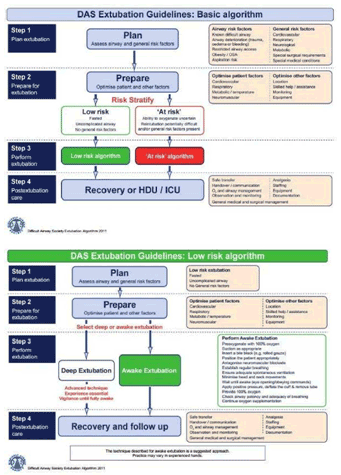
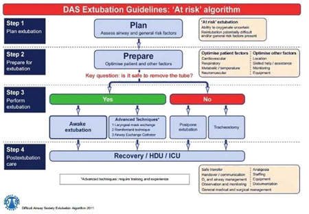
 
 
  Summary:
  Guidelines are useful in infrequent, life-threatening situations, and have been shown to improve outcomes. Several national guidelines for management of the airway have been published, but none has addressed extubation in detail. Extubation differs from intubation, in that it should always be an elective process with adequate time available to the anaesthetist for methodical management.
  Extubation practice is highly variable, and is not often formally addressed in training. Technical and nontechnical factors can contribute to adverse events at extubation but outcomes are improved by planning, organisation and communication.
  The DAS extubation guidelines promote the concept of an extubation strategy, involving a stepwise approach to planning, preparation and risk stratification, aimed at clear identification and management of patients ‘at risk’ during extubation.
  The evidence base for extubation practice is limited, so inevitably some of the recommendations in these guidelines are based on expert opinion.
  Awake extubation is the preferred technique for most patients. However, deep extubation, laryngeal mask exchange, remifentanil infusion and the use of airway exchange catheters may be beneficial in certain clinical situations.
  Delaying extubation or performing an elective tracheostomy should be considered when it is unsafe to extubate.