ISA KANYAKUMARI

ANAESTHESIA PEARLS


  KETAMINE IS GOING TO BE OUT OF REACH!
  The UN Commission on Narcotic Drugs will hold its 58th meeting in Vienna, starting on 9th March, and there will be an attempt to place further international restrictions on the distribution and use of ketamine.
  NAESTHETIC CONSIDERATION IN AIDS PATIENTS
  Fact Sheet on the Proposal to Discuss International Scheduling of Ketamine at the 58th CND
  Introduction
  Ketamine is an essential medicine used for anaesthesia. It is the only available anaesthetic for essential surgery in most rural areas of developing countries, home to more than 2 billion of the world’s people. Scheduling ketamine will leave these populations with no alternative anaesthesia for essential surgery, and will further deepen the already acute crisis of global surgery.
  The Commission on Narcotic Drugs, 58th Session, has been asked to review a proposal to place ketamine in Schedule I of the 1971 Convention (E/CN.7/2015/7 and E/CN.7/2015/81). As per the terms of the 1971 Convention on Psychotropic Substances, the WHO Expert Committee on Drug Dependence (ECDD) critically evaluated ketamine in 2006, 2012 and 2014. Based on accumulated evidence and data on non-medical use, diversion and trafficking, and evidence of ketamine’s therapeutic value, the ECDD does not recommend that the CND place ketamine under international control.
  A wide range of national and international civil society organizations, have voiced concern about the proposal. This basic fact sheet provides compelling legal, medical and social arguments against placing ketamine in any schedule of the 1971 Convention. Section I covers definitions and substantive issues; Section II, procedural issues.
  Section I: substantive issues
  What is ketamine? And how important is ketamine in human and veterinary medicine?
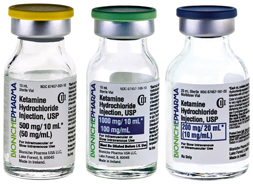
  Ketamine is a medication used as an anaesthetic in human and veterinary medicine. Because it is readily available, easy to use and inexpensive, ketamine is one of the most commonly used anaesthetic agents in developing countries. It is also used recreationally in some countries as a “party drug.” The World Health Organization considers ketamine an “essential medicine” and does not recommend scheduling it under the international substance control conventions. Regarding essential surgery, a WHO document states that ketamine must be accessible in all facilities where anaesthesia is needed, in order to ensure safe and affordable surgical care.
  Ketamine is also the primary anaesthetic used in veterinary practice. Scheduling ketamine would restrict access and lead to losses in the agricultural economy.
  How harmful is non-medical use of ketamine?
  The non-medical use of ketamine has limited harmful effects. After a notification by China suggesting that ketamine be placed in Schedule I of the 1971 Convention, the ECDD critically reviewed it again in 2014. The ECDD considered peer reviewed reports and data regarding ketamine’s recreational use in some urban areas, the likelihood of its potential to cause dependence if used non-medically, epidemiological evidence of morbidity and mortality rates, as well as records of police seizures of illicit supplies. After weighing all the reports, this international expert panel concluded that “ketamine abuse currently does not appear to pose a sufficient public health risk of global scale to warrant scheduling” and recommended that “ketamine not be placed under international control at this time”.
  Why does the WHO ECDD recommend against international control of ketamine?
  Further to the above, the ECDD considered evidence submitted from all over the world that ketamine is widely used as an anaesthetic in human and veterinary medicine, especially in low- and middle-income countries, as well as in emergency situations. Ketamine is easily administered by trained providers. Compared to anaesthetic gases, which require costly equipment and appropriately trained specialists, it is inexpensive and safe to administer. Since many countries have no appropriate or affordable alternatives, scheduling ketamine would force patients in those regions to forego lifesaving essential surgery, further compromising realization of the health-related Millennium Development Goals.
  What would be the public health impact of a placing ketamine in Schedule I?
  Under the terms of the 1971 Convention, medicines in Schedule I have “very limited medical usefulness”. Parties to the Convention are obliged to prohibit any medical use of a Schedule I substance except by "persons directly under control of the government,” and even use for and by those persons is very restricted (Art. 7). Providers in non-government institutions and clinicians in remote areas, especially in resource poor settings, will be unable to use ketamine if it is placed in Schedule I.
  The control stipulated for Schedule I substances would be very inappropriate for ketamine, given its critical use in developing countries and in zones of armed conflict where high-tech resources, labs, operating rooms etc. are unavailable.
  People living in rural areas of low-resource countries will not have access to essential surgery if ketamine is less available, or completely unavailable. International restrictions could potentially affect the health of an estimated 2 billion or more people, living mainly in Africa, Asia and Latin-America.
  What about placing ketamine in another schedule?
  The WHO ECDD did not recommend placing ketamine in any schedule. Hypothetically, however, placing ketamine in any other schedule would also limit its availability and accessibility, both of which are mandatory criteria for essential medicines. The cumulative evidence from over fifty years of international drug control shows that restrictive regulatory requirements imposed by scheduling essential medicines create costly burdens for low resource countries. Strict scheduling requirements result in decreased clinical use or abandonment altogether. In the words of the Convention, “the use of psychotropic substances for medical and scientific purposes is indispensable and […] their availability for such purposes should not be unduly restricted".
  Is there a way to limit misuse and diversion of ketamine?
  Ketamine is difficult to synthesize and does not lend itself to large scale illicit manufacture. UNODC reports that China is the source of much of the world's illicitly consumed ketamine, which has either been diverted from legitimate pharmaceutical manufacture, or illicitly produced and then domestically or internationally trafficked. The NGOs endorsing this letter call on the government of China to take the necessary steps to prevent the diversion, illicit manufacture and export of ketamine beyond its national frontiers, rather than to promote international scheduling.
  Section II. Procedural Issues
  Can the 58th CND decide to schedule ketamine under the 1971 Convention?
  Article 2, paragraph 5, of the 1971 Convention states that the Commission may add a substance to a schedule only following an explicit WHO recommendation to do so. As per the official Commentary to the Convention (p. 46): “the phrase ‘recommendations on control measures,’ means ‘recommendations on the schedule in which the substance in question should be placed”. Since the WHO recommended in 2014 that ketamine not be placed in any of the schedules, there is no extant recommendation that could legally serve as the basis of a scheduling decision by the CND.
  Under these circumstances, placing a scheduling proposal for ketamine on the CND agenda would contravene the terms of the 1971 Convention.
  Can the CND disregard a WHO recommendation to schedule or not to schedule a substance?
  No. As discussed above, the CND can consider scheduling a substance only if the WHO has recommended that the substance be placed in a schedule. In such a case, State Parties can select a schedule other than the schedule recommended by WHO based on relevant economic, social, administrative or other factors. Article 2, paragraph 5 of the 1971 Convention states, however, that the WHO’s medical and scientific determination is final.
  Notifications by Parties to the Convention are not a basis for CND decision making about including substances in the treaty schedules. Regarding medical and scientific aspects, these notifications can only serve to inform other Parties, the CND, and WHO for the purposes of their evaluative process.
  Following notifications by State Parties, CND can only decide to take provisional control measures, while awaiting the recommendation of the WHO. But in the case of ketamine, since WHO already issued its recommendation, the option of a provisional control measure no longer applies.
  How does the continuing pressure on WHO affect its ability to fulfil its mandate under the international substance control conventions?
  Following requests from China, the International Narcotics Control Board and others, the World Health Organization’s ECDD reviewed ketamine in 2006, 2012 and 2014, each time concluding that scheduling is not warranted. Ketamine’s medical importance has been well documented, while evidence of its harmful properties is limited. Performing additional reviews in the near future would be inefficient and costly. WHO is also under pressure to evaluate a large number of New Psychoactive Substances (NPS) and to re-evaluate several scheduled substances it had evaluated in the distant past. WHO’s substance evaluation mandate gives it a critical role in safeguarding global health. The ability to fulfil this mandate is dependent on the States Parties and UN bodies abiding by the conventions – in this case, following the WHO’s expert recommendations.
  Note:
  China, a major producer of ketamine, is understood to be concerned by its abuse and it has the support of a number of other countries, including Russia. It will need backing from two-thirds of the 53 member states. The UK has said it will abstain on the vote, which is tantamount to a no.
  Campaigners argue the proposal should not have been brought at all. “Legally, WHO [World Health Organisation] must consent before the commission can add any drug to schedule 1,” said Amir Attaran, law professor at the University of Ottawa in Canada. “WHO has thrice come out unequivocally against scheduling ketamine.”
  He argues that the UN Office on Drugs and Crime, the secretariat of the Commission on Narcotic Drugs, should not have put the draft resolution forward for discussion.
  “China has been pushing for a long time,” said Willem Scholten, an independent consultant in controlled medicines in the Netherlands who was secretary of WHO’s expert committee on drug dependence until two years ago. Beijing first put it forward in 2005 and it was reviewed again in 2012 and in 2014 – each time WHO advised against it because of the potential impact on health in the developing world.
  When the resolution is debated by the commission, it is understood that the scientific and medical opinion of the WHO will not be challenged. The argument then will centre on the experiences of crime and drug abuse put forward by the individual member states.
  SOURCE:
  The Gaudian and Message from AAGBI President, Dr Andrew Hartle
  The myth of the difficult airway
  For years, anaesthetists have tried to predict the difficult airway using various clinical signs and prediction models. In this issue of Anaesthesia, Nørskov et al. present a study of a large cohort of 188 064 patients in Denmark and come to a disappointing conclusion: we are not good at it. Of 3391 difficult intubations, 3154 (93%) were unanticipated. When difficult intubation was anticipated, only 229/929 (25%) had an actual difficult intubation. Difficult mask ventilation was unanticipated in 808/857 (94%) cases.
  NAESTHETIC CONSIDERATION IN AIDS PATIENTS
  The incidence and the definition of the difficult airway, difficult laryngoscopy and difficult intubation are not well defined.
  Cook and MacDougall-Davis recently summarised that CICO has an incidence of 1:50 000 and failed intubation occurs in 1:2000 elective cases, but up to 1:200 in emergencies. Rocke et al. reported difficult intubation in 7.9%, and very difficult intubation in 2%, of parturients undergoing general anaesthesia for caesarean section. In a mixed surgical population, Rose et al. noted that 2.5% of patients required two laryngoscopies to achieve tracheal intubation and that 1.8% required more than three. This suggests that difficulty with intubation occurs more frequently during obstetric anaesthesia, but that the frequency of very difficult intubation is similar in obstetric and non-obstetric surgical populations. Further, that these numbers haven't changed over the years.
  Furthermore, if we use the well-known definition for the difficult airway endorsed by the American Society of Anesthesiologists: “a clinical situation in which a conventionally trained anaesthesiologist experiences difficulty with face mask ventilation of the upper airway, difficulty with tracheal intubation or both”, it will unfortunately not make a clear difference for clinical practice. A global competence profile of the ‘conventionally trained anaesthesiologist’ does not exist and according to the definition, the clinician can only find out whether a patient has a ‘difficult airway’ after intubation has failed.
  Prediction models
  Using a three-tier classification, Mallampati et al. reported difficult direct laryngoscopy in the majority of patients with a poor view of the pharyngeal structures. Samsoon and Young reviewed a series of patients with known difficult intubation and added a fourth class (no pharyngeal structures seen): in patients with difficult laryngoscopy, classes 3 and 4 predominated. Unfortunately, subsequent evaluations showed that Mallampati/modified Mallampati scores poorly predict difficult intubation.
  Wilson et al.'s model, based on grading patients’ weight, head and neck movement, jaw movement, mandibular size and prominence of the upper incisors, predicted difficult intubation with sensitivity of 75% and specificity of 88%.
  In parturients undergoing caesarean section, Rocke et al. utilised the modified Mallampati classification combined with other characteristics (short neck, obesity, missing/protruding maxillary incisors, single maxillary tooth, facial oedema, swollen tongue and receding mandible)
  An easy, first-attempt intubation occurred in 96% of class-1 airways, 91% of class-2, 82% of class-3 and 76% of class-4 airways. Surprisingly, most class-4 airways were not difficult to intubate, and only 4-6% of class-3/4 airways were considered to be very difficult intubations. The following emerged as aetiological factors predicting difficult or failed intubation: airway class 2 (RR 3.23); airway class 3 (RR 7.58); airway class 4 (RR 11.30); short neck (RR 5.01); receding mandible (RR 9.71); and protruding maxillary incisors (RR 8.0). Obesity and a short neck were linked factors, with obesity being eliminated as a risk factor if short neck was excluded.
  While it can be concluded that airway management is a highly complex procedure, the ‘difficult airway’ does not exist.
  A previously healthy patient, whose airway is scored as Mallampati class 1 and whose trachea can be intubated with basic airway management skills, may become ‘difficult’ when he presents in septic shock and with a low oxygen saturation, to an emergency physician in a remote hospital who performs only ten tracheal intubations a year.
  In contrast, a Mallampati class-4 airway can represent a routine intubation for an anaesthetist experienced in awake intubation, even after major head and neck surgery with free flap reconstruction. For these doctors, the definition of a difficult airway will be different, and accurate prediction of intubation problems is impossible with current methods.
  Most of our patients will have basic airways. In the rare event of unexpected difficulties, it should be possible to call for help early and/or to wake up the patient, without undue risk.
  The advanced airway
  In patients with advanced airways, it must be anticipated that airway management can be challenging, because of the presence of one or more of the complexity factors. In these cases, special measurements or advanced skills may be needed, for example the immediate availability of a dedicated airway management trolley or, in cases with many complexity factors, the help of an airway management expert and/or head and neck surgeon.
  In the 4th National Audit Project (NAP4), the following complexity factors were strong predictors for complications: body mass index (Patient factors); head and neck conditions (Patient factors); absence of capnography (Equipment factors); communication problems (Human factors); out of theatre location (Location); and wrong emergency surgical technique (Experience, Time pressure)
  The term ‘difficult airway’ has been a cause of confusion and that we should start redefining airway assessment and management.
  SOURCE:
  The myth of the difficult airway: airway management revisited. Anaesthesia, Volume 70, Issue 3, pages 244–249, March 2015.
  Ultrasonography Basics
  INTRODUCTION
  Ultrasound imaging is a diagnostic medical procedure that uses high-frequency sound waves to produce dynamic images (sonograms) of organs, tissues or blood flow inside the body. The frequency of an ultrasound wave is above 20,000 Hz and that of medical ultrasound is 2.5-15 MHz. Human hearing is in the 20-20,000 Hz range. The speed of sound varies in different biological media, however, the average value is assumed to be 1,540 m/sec for most human soft tissues. The speed of sound can be calculated by multiplying wavelength to frequency. Thus sound with a high frequency has a short wavelength and vice versa.
  Ultrasound wave creation
  An ultrasound wave is generated when an electric field is applied to an array of piezoelectric crystals located on the transducer surface. Electrical stimulation causes mechanical distortion of the crystals resulting in vibration and production of sound waves. The sound waves emitted from the transducer are transmitted into the body, reflected off the tissue interface and returned to the transducer. These are then converted into an electric signal, which is processed and displayed as an image on the screen. The conversion of sound to electrical energy is called the piezoelectric effect.
  Transducer selection
  The first step in the systematic scanning process is to select an appropriate transducer. This depends upon the depth and the location of the area to be scanned.
 
  • High frequency (7-15 mHz) (linear) transducer has higher resolution but poor penetration. It is useful for peripheral nerve blocks and central venous cannulation as these structures are superficially located.
  • Low frequency (2-5 mHz) (curved array) transducer has lower resolution but better penetration. This is useful for epidural/spinal blocks as these structures are deeply situated.
  An orientation marker is usually located on the side of the ultrasound transducer. It helps to orient the image on the screen with respect to the patient position. (Figure 1)
  Figure 1. Different types of transducers. Linear (left); Curved (right). The orientation marker on the linear transducer is marked with a circle.
  NAESTHETIC CONSIDERATION IN AIDS PATIENTS
  Ultrasound imaging echogenicities
  Different structures emit different signals on ultrasound imaging, which are termed as echogenicities. Hyperechoic structures appear white and bright on the ultrasound image, while isoechoic have the same density as the surrounding structures and appear grey. Hypoechoic structures appear dark, black and produce a weak signal.(Figure 2)
  Figure 2. Examples of different ultrasound echogenicities.
  NAESTHETIC CONSIDERATION IN AIDS PATIENTS
  Identification of structures
  Different structures can be identified based on their anatomical location, shape and echogenic pattern. The examples of blood vessels, nerves, bones and ligaments are given below:
 
Blood vessels: Veins and arteries, in the transverse view, appear as circular hypoechoic structures surrounded by hyperechoic rims. Arteries can be easily identified by their pulsatile nature. Veins collapse with the pressure of the ultrasound probe while the arteries remain stiff. (Figure 3)
Figure 3: Top: Internal jugular vein (IJV) and carotid artery (CA) in the transverse view with the transducer held on the skin without pressure (left) and with pressure (right). Note that with pressure, IJV appears compressed while the CA has retained its shape. Bottom: These blood vessels in the longitudinal plane.
  NAESTHETIC CONSIDERATION IN AIDS PATIENTS
 
Nerves: Nerves below the clavicle and in the lower limbs are predominantly hyperechoic and have a honey comb appearance. The degree of hyperechogenicity likely reflects the amount of connective tissue within the nerve. In the longitudinal view, they appear as hypoechoic longitudinal elements (fascicle groups) interspersed with hyperechoic perineural connective tissues. (Figure 4)
Figure 4: Top: Interscalene brachial plexus in transverse view. Bottom: A nerve in longitudinal plane. Note the honeycomb appearance of nerves represented by arrowheads in both planes. (SCM=sternocleidomastoid; ASM=anterior scalene muscle; MSM=middle scalene muscles).
  NAESTHETIC CONSIDERATION IN AIDS PATIENTS
 
Ligaments and Bones: These appear hyperechoic on the ultrasound image. However, bone has high acoustic impedance and does not allow the sound waves to pass through it. This produces a hypoechoic shadow underneath intensely hyperechoic bony structure, termed as Acoustic shadow. Hence while scanning through bony structures, it is necessary to find Acoustic window, an area through which sound waves can be transmitted to visualize the deeper structures. (Figure 5)
Figure 5: Left: Ultrasound view of the lumbar spine in transverse plane. Right: Ultrasound image of the lumbar spine in longitudinal plane with acoustic windows between lamina and acoustic shadows under lamina. (LF=ligamentum flavum; VB=vertebral body; AP=articulate process; TP=transverse process; La=lamina).
  NAESTHETIC CONSIDERATION IN AIDS PATIENTS
  Scanning planes
  Scanning in different planes or axes helps in identification of the structures more accurately and facilitates the location and needling of the target areas. Longitudinal axis usually refers to scanning in a plane parallel to the structures while transverse axis refers to scanning in a plane perpendicular to the structures (Figure 6).
  Figure 6. Scanning in longitudinal (left) and transverse (right) planes.
  NAESTHETIC CONSIDERATION IN AIDS PATIENTS
  Image optimization
  Fine movements of the transducer can help in obtaining the best image.
 
Depth settings: Adjust the depth of the image for optimal resolution so that the area of interest is centered on the screen. For scanning for the superficial nerve blocks, set the depth up to 5 cm, while for neuraxial scanning, set it up to 13 cm.
Gain settings: Adjust gain settings to allow amplification or reduction of intensity of echo. a) Increased gain=brighter image display; b) Decreased gain=darker image display. You can change the gain settings of the entire image (overall) or of any particular field such as near field, or far field. Usually setting the gain to "optimum", provides an appropriate image display.
  Needle alignment
 
In-plane needle alignment (longitudinal, long-axis): In-plane needle alignment refers to aligning the needle with the long-axis of the transducer (along the ultrasound beam) so that the entire shaft and tip of the needle are visible. One of the disadvantages of the in-plane needle view is that, it is easy to lose the image with a slight movement of the transducer as the ultrasound beam is thin. This technique requires excellent hand-eye coordination. It is a preferred technique for Transversus Abdominus Plane block (Figure 7).
Out-of-plane alignment (transverse or short axis): This refers to when the transducer and the needle are perpendicular to each other. It is important to slide the transducer along the shaft of the needle to identify the needle tip. Both the needle tip and shaft in cross section appear as a hyperechoic white dot on the screen. Since only the needle tip is observed as a bright dot, it is sometimes difficult to accurately observe the needle during advancement. Despite this, it is an easier approach for peripheral nerve blocks and central venous cannulation (Figure 7).
  Figure 7: In in-plane technique (left), needle is aligned in the plane of thin ultrasound beam allowing the visualization of the entire shaft and the tip. In out-of plane technique (right), the ultrasound beam transects the needle, and the needle tip or the shaft is observed as a bright spot in the image.
  NAESTHETIC CONSIDERATION IN AIDS PATIENTS
  SOURCE:
  Dr. Rafeek Mikhael MD, PhD, Department of Anesthesia, Toronto General Hospital, University of Toronto.
  Mount Sinai Hospital, Department of Anesthesia, Perioperative Interactive Education. (PIE)