| NAP4 - 4th National Audit Project of The Royal College of Anaesthetists |
| |
Major complications of airway management in the United Kingdom |
| |
 |
 |
 |
 |
 |
| |
The 4th National Audit Project of the Royal College of Anaesthetists and the Difficult Airway Society (NAP4) was designed to answer the questions; |
| |
 |
What types of airway device are used during anaesthesia and how often? |
|
How often do major complications, leading to serious harm, occur in association with airway management in anaesthesia, in the intensive care units and in the emergency departments of the UK? |
|
What is the nature of these events and what can we learn from them, in order to reduce their frequency and consequences? |
|
| |
Recommendations at a glance: |
| |
 Events at induction and during maintenance of anaesthesia
(excluding head and neck) (Chapter 7) Events at induction and during maintenance of anaesthesia
(excluding head and neck) (Chapter 7) |
| |
|
All anaesthetic departments should have an explicit policy for management of difficult or failed intubation (e.g. formal adoption of the Difficult Airway Society guidelines as departmental policy). The strategy should limit the number of intubation attempts. |
 |
|
Where difficulty with airway management is anticipated or has occurred previously a comprehensive airway strategy must be planned before induction of anaesthesia. Plans B and C should be discussed and the equipment and skills to carry them out must be available. |
|
| |
 |
|
Anaesthetic departments should provide a service where the skills and equipment are available to deliver awake fibreoptic intubation whenever necessary. |
|
If a flat capnograph is seen after attempted tracheal intubation the anaesthetist should actively exclude oesophageal intubation (and absolute airway obstruction). |
|
| |
|
Anaesthetists must assess all patients for risk of aspiration prior to anaesthesia. This applies particularly to urgent and emergency surgery. |
 |
|
Once placed a tracheal tube offers the highest protection against aspiration. |
|
Second generation SAD s may offer better protection than first generation devices but further research is needed to confirm and quantify this. |
|
Where facemask or laryngeal mask anaesthesia is complicated by failed ventilation and increasing hypoxia the anaesthetist should consider early administration of further anaesthetic agent and or a muscle relaxant to exclude and treat laryngospasm. |
 |
|
No anaesthetist should allow airway obstruction and hypoxia to develop to the stage where an emergency surgical airway is necessary without having administered a muscle relaxant. |
|
| |
Events at the end of anaesthesia and during recovery from anaesthesia (Chapter 8) |
| |
|
Patients should be assessed and optimised before tracheal extubation to ensure they are extubated with effective neuromuscular function, after preoxygenation and appropriate airway toilet. |
 |
|
Patients at high-risk of airway problems at emergence require a specific extubation and reintubationplan. Extubation should usually take place in theatre with the team assembled and may include the use of specific techniques to facilitate re-intubation. |
|
Supplementary oxygen is needed for transport after general anaesthesia and supplementary monitoring should be considered as recommended by the AAG BI. |
|
| |
|
In patients at risk of airway problems, an airway management plan should be conveyed to recovery staff which should include:
(1) potential problems,
(2) signs indicating concern,
(3) planned management,
(4) equipment required, and
(5)location of appropriate medical help if needed. |
|
| |
 |
|
Recovery room staff should be trained to an agreed standard in all hospital sites, this must include the prevention, early recognition and management of airway obstruction. |
|
Capnography has the potential to aid early detection of airway obstruction. It should be available and used in high-risk cases. |
|
| |
|
Tracheal tube and SAD obstruction by the patient biting should be prevented by the insertion of a bite block, an oropharyngeal airway, or the use of SAD s with an integral bite block. |
|
A full range of difficult airway equipment and experienced staff should be readily accessible in recovery. |
|
Patients who have potential airway problems or have had complications should be reassessed by the responsible anaesthetist before discharge. |
|
| |
CICV, emergency surgical airways and cricothyroidotomies (Chapter 13) |
| |
|
Patients with airway tumours are at high-risk of CIC V. In patients with symptoms of airway obstruction, airway imaging and nasendoscopy should be considered a minimum level of investigation in helping assess the options for anaesthetic airway management. Only in exceptional cases should anaesthesia proceed without this level of airway assessment. |
 |
|
| |
|
Securing the airway before induction of anaesthesia (by awake intubation or awake tracheostomy) should be considered in all cases where the airway is at risk from the presenting condition or where difficulty has been experienced previously. |
|
Where difficulty with airway management is anticipated or has occurred previously a comprehensive airway strategy must be in place before induction of anaesthesia. Plans B, C and D should be discussed with the team and the equipment and skills to carry them out must be available. |
|
All anaesthetic departments should provide a service where the skills and equipment are available to deliver awake fibreoptic intubation when it is indicated. |
|
Where there is a high suspicion that a cricothyroidotomy might be needed to rescue the airway, consideration should be given to placing this (as a needle or surgical procedure) prior to anaesthesia. |
|
All anaesthetists should be made aware of published guidelines and trained in their use. Unlimited attempts at intubation are not indicated. |
|
| |
 |
|
Even if it was not part of the initial airway management strategy, if CIC V occurs and waking the patient up is not an option, a muscle relaxant should be given before determining the need to proceed to a surgical airway. |
|
In patients considered to be at low-risk of aspiration who have other factors that mean that use of a SAD is at the limits of normality (e.g. patient position, access to the airway, patient size) consideration should be given to use of a second generation SAD . |
|
| |
|
In view of the above recommendations, and the frequency of these circumstances, it is recommended that all hospitals have second generation SAD s available for both routine use and rescue airway management. |
|
| |
Tracheal tube-related cases (Chapter 12) |
| |
|
Airway assessment should be performed and documented prior to anaesthetising a patient. |
 |
|
Plans for difficult or failed intubation should be made before induction of anaesthesia and should include the use of different devices both for direct laryngoscopy (e.g. alternative blades) and airway rescue (e.g. supraglottic airway devices). |
|
All anaesthetic departments should have an explicit policy for management of difficult or failed intubation (e.g. formal adoption of the Difficult Airway Society guidelines as departmental policy). |
|
| |
|
Rescue techniques that involve direct access to the trachea should be included in the policy for management of difficult or failed intubation. These techniques should be taught and practised with equipment that is available at that hospital. |
|
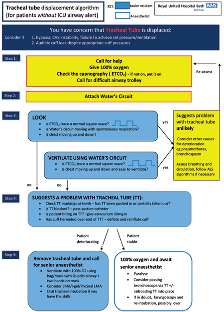
Capnography should be used during all intubations, irrespective of the location. |
|
Training of all clinical staff who may intubate patients should include interpretation of capnography. Teaching should include recognition of the abnormal (but not flat) capnograph trace during low cardiac output states and during cardiopulmonary resuscitation. |
|
All cases, but particularly those undergoing emergency surgery, should be assessed for risk of regurgitation and aspiration. |
|
On balance, rapid sequence induction should continue to be taught as a standard technique for protection of the airway. Further focused research might usefully be performed to explore its efficacy, limitations and also explore the consequences of its omission. |
|
Techniques that reduce the need for intubation involving blind placement of a bougie or introducer probably lessen the risk of trauma. Fibreoptic intubation and indirect laryngoscopy (e.g. videolaryngoscopes) may have a role. Further research is required. |
|
All anaesthetists should be trained in low-skill rescue intubation through a supraglottic airway. A technique using the Aintree Intubation Catheter is recommended. |
|
Fibreoptic endoscopy should be immediately available to confirm airway device placement in situations where capnography may be misinterpreted. |
| |
 |
 |
|
| |
Fibreoptic intubation (Chapter 14) |
| |
|
All anaesthetic departments should provide a service where the skills and equipment are available to deliver awake fibreoptic intubation whenever it is indicated. |
 |
|
Where FOI is considered the optimal method of securing the airway, an awake technique should be considered unless contraindicated. |
|
Fibreoptic intubation is most effective in co-operative patients. Airway patency and co-operation may be lost by over-sedation. Where complex sedation techniques are to be used strong consideration should be given to delegating the provision of sedation to an anaesthetist not performing the tracheal intubation. |
|
| |
|
Following awake fibreoptic intubation, general anaesthesia should only be induced after the tracheal tube has been railroaded, its position checked and the cuff inflated to seal the airway. |
|
AFOI may fail. A back-up plan should always be worked out in advance |
|
Oral fibreoptic intubation should be taught and practised alongside nasal fibreoptic intubation so that it can be considered in patients in whom nasotracheal intubation is not specifically indicated. |
|
If no additional investigations have been performed (or performed recently) consideration should be given to awake flexible nasendoscopy in the operating theatre to reassess the situation prior to starting anaesthesia. |
|
Successful management of these cases requires not one plan but a series of plans pre-formulated into an ‘airway management strategy’. This strategy should be agreed by the anaesthetist and surgeon prior to starting. The theatre team should be briefed on the strategy and the necessary equipment and personnel assembled. |
|
The anaesthetic management of any case which may involve surgical tracheostomy as a rescue technique should start in the operating theatre. Consideration should be given to anaesthetising all complex head and neck cases in the operating theatre. |
|
Multiple attempts at direct laryngoscopy in patients with head and neck pathology should be avoided. |
|
When patient factors make fibreoptic intubation the preferred option in patients with head and neck pathology, consideration should first be made to performing it awake. The airway strategy should accept it may fail, particularly when performed in an unconscious patient. |
|
When inhalational induction is the primary plan for cases involving head and neck pathology the airway strategy should accept it may fail with loss of the airway. A clear rescue plan, that does not assume the patient will wake, should be in place before anaesthesia starts. |
|
When emergency cricothyroidotomy is included as part of the airway strategy for cases involving head and neck pathology success should not be assumed. The airway strategy should accept it may fail. |
|
| |
 |
|
Anaesthetic management of these patients is predictably difficult and difficulty may affect all approaches to the airway. Senior anaesthetists and surgeons must be involved. While opening wounds to relieve haematoma may reduce airway compression it will not resolve resultant airway oedema and the airway is likely to remain difficult to manage. |
|
| |
|
For cases with head and neck pathology the team managing the patient should not disperse until the patient is clearly managing their own airway and is safe. |
|
A flexible fibrescope should be immediately available on the IC U to check position of tracheal/ tracheostomy tubes and assist with fibreoptic intubation or percutaneous tracheostomy placement. |
|
Clear lines of communication are required between the various teams that manage airway problems related to tracheostomy (IC U, anaesthetic and ENT clinicians) in order to best manage such patients with potentially difficult airways. Mechanisms are also required within teams so senior staff are appropriately available and involved when adverse airway incidents occur. |
|
| |
Airway assessment and planning (Chapter 17) |
| |
|
All patients should have an airway assessment performed and recorded before anaesthesia. This involves bedside interactive tests. |
|
All patients should have their risk of aspiration assessed and recorded before anaesthesia. The airway management strategy should be consistent with the identified risk of aspiration. |
|
Awake intubation should be used when it is indicated. This requires that anaesthetic departments and individual anaesthetists ensure such a service is readily available. |
|
All anaesthetic departments should have an explicit policy for management of difficult or failed intubation (e.g. formal adoption of the Difficult Airway Society guidelines as departmental policy) and for other airway emergencies. Individual anaesthetists should use such strategies in their daily practice. |
| |
 |
 |
 |
|
| |
Obesity (Chapter 20) |
| |
|
Hospital management need to be aware of the additional time and resources required to safely anaesthetise obese patients. |
 |
|
Provision must be made for anaesthetists to evaluate obese patients before surgery. Morbidly obese patients and obese patients with significant morbidity should be formally assessed by an anaesthetist in a setting without time limitations. |
|
| |
|
Obese patients require thorough preoperative evaluation of co-morbidities. Evidence of OSA should be sought routinely. |
|
Airway assessment should form part of the evaluation of all obese patients and should include an evaluation of possible rescue techniques. |
|
Awake intubation should be considered in those patients in whom it would be difficult to establish rescue oxygenation or emergency surgical airway (e.g. those obese patients in whom the cricothyroid membrane or trachea cannot be identified). |
|
If AFOI is chosen, extreme care is required in titration of sedatives and monitoring, in order to avoid airway obstruction and periods of apnoea. |
|
| |
 |
|
Failure of regional anaesthesia may necessitate general anaesthesia. Obese patients undergoing regional anaesthesia still require a strategy for airway management. Regional anaesthetic blocks should be thoroughly checked before surgery. All theatre staff must be aware of the hazards posed by intra-operative conversion from regional to general anaesthesia. |
|
| |
|
Pre-oxygenation, performed to high standards, should be used for all obese patients prior to general anaesthesia. |
|
Organisations and individual anaesthetists should procure and use airway devices and techniques that meet the specific needs of obese patients. Safety should take priority in the decisions made. |
|
The maintenance of a clear airway in patients admitted to IC U requires continuous preparedness for insertion of a tracheal tube or tracheostomy in difficult circumstances. As in theatre this requires an airway strategy (ability to recognise and diagnose the problem, the right equipment and personnel to respond with a series of pre-formulated, logical and sequential plans). |
|
| |
Aspiration of gastric contents (Chapter 19) |
| |
|
Anaesthetists must assess all patients for risk of aspiration prior to anaesthesia. This applies particularly to urgent and emergency surgery. Where significant doubt exists, the higher risk should be assumed. |
 |
|
The airway management strategy should be consistent with the identified risk of aspiration. Where reasonable doubt exists it is likely to be safer to assume increased risk and plan accordingly. |
|
No matter how low the perceived risk of aspiration, when anaesthesia is induced, the equipment and skills should exist to detect, and promptly manage, regurgitation and aspiration. |
|
On balance, rapid sequence induction should continue to be taught as a standard technique for protection of the airway. Further focused research might usefully be performed to explore its efficacy, limitations and also explore the consequences of its omission. |
|
To maximise the likelihood of good quality cricoid force being applied, those who perform cricoid force should be trained in its methodology, should practise at regular intervals and should consider the use of simple methods of simulation. |
|
If tracheal intubation is not considered to be indicated but there is some (small) increase or concerns about regurgitation risk a second generation supraglottic airway is a more logical choice than a first generation one. |
|
Where aspiration has been recognised as a risk at induction, steps should be taken to reduce the risk of aspiration at emergence. |
|
Anaesthetists caring for patients undergoing intra-oral surgery should be educated in the prevention, detection and management of blood clot aspiration. |
|
| |
Obstetrics (Chapter 22) |
| |
|
 |
 |
|
Despite the relative infrequency of general anaesthesia for caesarean section, obstetric anaesthetists need to maintain their airway skills including strategies to manage difficult intubation, failed intubation and CIC V. |
|
Obstetric anaesthetists should be familiar and skilled with supraglottic airway devices for rescuing the airway: particularly those designed to protect from aspiration and to facilitate ventilation and or intubation. |
|
A flexible fibrescope may have several roles in the obstetric setting. Anaesthetic departments should provide a service where the skills and equipment are available to deliver awake fibreoptic intubation whenever it is indicated. |
|
All staff working in the recovery area of a delivery suite including midwifery staff must be competency trained. Skills must be regularly updated. |
|
| |
Appendices: |
| |
 |
| |
 |
| |
 |
| |
Source: |
| |
4th National Audit Project (NAP4), Report and findings March 2011. |