Conference Lectures
Peripheral nerve injuries associated with anaesthesia
Dr Ashok V Deshpande Intensivist
Bharati Medical College and Hospital Sangli
Peri-operative nerve lesions can mar a successful surgical procedure, handicap a patient with a severe functional disability and leave the medical team facing possible protracted and unpleasant litigation. The true incidence of peri-operative nerve damage remains unclear and, as a complication, it is probably under-reported. The presenting neurological symptoms may appear minor in nature to the attending physician, when compared with the clinical disorder that led to the initial surgical procedure, and, as such, may be dismissed. There is a belief that the lesion will be self-limiting with a full and spontaneous recovery; however, this is often not the case .It is our impression that the incidence of these lesions is sufficiently great to warrant further study into their causation and prevention.
Nerve palsies following anaesthesia have been observed since shortly after the introduction of ether .Early writers blamed toxic properties of the anaesthetic agent, but Budinger was the first to recognise that the majority were secondary to malpositioning of the patient on the operating table with consequent stretching and/or compression of nerves . The ulnar nerve , postoperative ulnar neuropathy comprised one third of the injuries, the brachial plexus nerves 23% and lumbosacral roots 16% .Although the mechanism of injury is uncertain, it is often suspected to be excessive pressure or undue stretching of the nerve.The nerve palsies associated with general anaesthesia,75% are due to medical negligence. The commonest nerve injuries are ulnar nerve palsies and common peroneal nerve injuries
Aetiology
There are many possible ways in which nerves can be damaged in the peri-operative phase. Events such as direct injury by needles, instruments, suturing, or the injection of neurotoxic material, or even thermal insults from diathermy, can cause nerve damage. The relationship between the agent and its effect is usually unclear. Less obvious, but more frequent adverse events, involve mechanical factors such as compression, stretch angulation, percussion or transection. Ischaemia is a crucial element in many of these injuries The interdependence between mechanical and ischaemic factors is well recognised, but which is the more important factor in individual cases remains controversial.
Predisposing factors
Nerves may be unduly susceptible to trauma as a result of a pre-existing generalised peripheral neuropathy or a local compression neuropathy, whether overt or subclinical, or rarely as a hereditary predisposition. Nerve lesions are more common in diabetic patients than in the general population and, indeed, the development of a peri-operative nerve lesion should be an indication for further investigation of a possible underlying disease process. Patients who develop a postoperative ulnar neuropathy on one side often have abnormal ulnar nerve conduction on the other side Other structural causes include congenital abnormalities in the region of the thoracic outlet and arthritis or instability of the elbow joint. Superficial nerves, such as the ulnar and common peroneal nerves, are especially vulnerable in patients who are thin . Among the systemic factors potentially involved in the pathogenesisofperi-operative nerve lesions are hypovolaemia dehydration, hypotension, hypoxia and electrolyte disturbances. A high incidence of postoperative neuropathies has been reported after induced hypothermia .
Surgical factors
The surgical procedure itself is relevant. All surgical disciplines employ patient positioning techniques, which may predispose to particular nerve injuries.General surgical procedures on the lower abdominal or inguinal region may give rise to damage to the ilioinguinal, iliohypogastric or genito-femoral nerves either as a result of direct trauma or from excessive flexion of the thigh onto the abdomen. Post vasectomy neuralgia affecting the scrotum occurs as a result of damage to the ilioinguinal nerve. The LloydDavies position for rectal or anal surgery, or the exaggerated lithotomy position for radical prostate surgery both carry a particularly high risk because the patient's legs are flexed and abducted in supports for a prolonged period. During cardiac surgery, median sternotomy is associated with a relatively high risk of peri-operative injury to nerves of the brachial plexus.Possible causes include traction on the brachial plexus, compression of its medial cord by the first rib, or fracture of the first rib leading to a hyperabduction thoracic outlet compression syndrome. Coronary artery bypass surgery has also been associated with a high incidence of postoperative ulnar neuropathies
The sciatic, femoral or obturator nerves may be damaged during total hip arthroplasty, the risk being higher in female patients and in operations requiring significant lengthening of the extremity stretching the sciatic nerve.
The harvesting of bone or vein grafts may be followed by damage to nerves in the vicinity of the donor site. For example, meralgiaparaesthetica caused by injury to the lateral femoral cutaneous nerve has been reported following iliac bone procurement .Neurosurgery and the use of the sitting position for approaches to the posterior fossa is associated with nerve injuries. The common peroneal nerve is frequently involved. Recurrent laryngeal nerve injury has been described after the use of transoesophageal echocardiography in two patients who underwent craniotomy in the sitting position. It is possible that the large size of the probe, tracheal intubation and excessive neck flexion resulted in pressure on the laryngeal nerve .
Anaesthetic factors
Peripheral nerve damage is associated with regional anaesthesia and nerve damage as a result of direct needle trauma and, although relatively rare, is avoidable with a meticulous technique. Peripheral nerve damage is not as dramatic as central neural damage, which may occur with spinal or extradural techniques. Neurological damage may only become apparent up to 1 week after the procedure. In normal clinical practice, an anaesthesiologist may not see patients after a nerve block, when the neurological signs will have become manifest .The type of needle used in performing a regional technique has been a topic of debate for many years. Needle trauma may damage neural blood vessels causing extra- or intraneuralhaematoma, and possibly direct trauma to neural fascicles. Either of these may result in adiscontinuity of fibres and subsequent pain. An immobile needle technique as described by Winnie is still to be recommended. However, the debate as to whether to eliciteparaesthesia or not is still contentious. The use of blunt atraumatic block needles has been suggested because they may help in identifying tissue planes
Local anaesthetics used in clinical concentrations are not neurotoxic when applied extraneurally. There may be differences in neurotoxicity in substances injected intraneurally and this has been demonstrated with epinephrine . It is theoretically possible that any preservative in a solution could be neurotoxic and it would therefore be wise to use preservative-free solutions for nerve blocks. The introduction of chemical agents used in skin preparation is a theoretical complication and has not been implicated in peripheral nerve damage. The use of pneumatic tourniquets, to produce a bloodless field, has greatly reduced the risk of nerve injury as a result of better control of the compressive pressure. There is still a significant risk of compression nerve damage and tourniquet time should be kept as short as possible. Lingual nerve damage has been associated with tracheal intubation and the use of the laryngeal mask. Irregular and over inflation of the cuff and nitrous oxide distension of an air-containing cuff are factors thought to be contributory.
Classification of nerve injury
The degree to which a nerve is damaged has implications with respect to its function and potential recovery. There are essentially two general classification systems.
Seddon's classification
Sunderland's classification
Seddon's classification describes three groups:
1. Neurapraxia 2. Axonotmesis 3.Neurotmesis.
Sunderland's classification describes five types of injury and depends exclusively upon which
connective tissue components are disrupted
Neurapraxia describes a mild degree of neural insult that results in impulse conduction failure across the affected segment. It is reversible.
Axontomesis occurs when only the axon is physically disrupted with preservation of the endoneurial and other supporting connective tissue structures. Recovery of function
depends upon time for the process of Wallerian degeneration and neural regeneration to occur.
Neurotmesis is the greatest degree of disruption a nerve can incur and is complete disruption of all supporting connective tissue structures. The nerve is completely severed and there is no continuity and this carries a very poor prognosis for complete functional recovery.
Clinically, the prognosis for spontaneous recovery in Sunderland's is
Type 1 good
Type 2 fairly good
In the remaining groups the prognosis is poor,
Type 3 surgery may /may not required
Type 4 surgery required
Type 5 Surgery required
Specific forms of nerve injury
Brachial plexus
These are most frequently related to excessive stretching of the cord. In general terms, stretch of the brachial plexus is particularly induced by arm abduction, external rotation and posterior shoulder displacement . Circumstances in which this may occur include extension and lateral flexion of the head to one side with the patient supine, and then abduction, external rotation and extension of the arm by allowing it to drop away from the side of the body. Extreme abduction of the arm so that the hand rests above the head also causes considerable stretch on the brachial plexus roots .Compression of the brachial plexus can also be implicated in brachial plexus injury. This has been described with upward movement of the clavicle secondary to sternal retraction and median sternotomy. Compression also plays a predominant role in injury with the patient in the lateral decubitus position when the plexus is compressed against the thorax by the humeral head. Anatomical variation of the thoracic outlet, in particular the presence of an extra rib on the seventh cervical vertebra, may provide a predisposing cause . Brachial plexus lesions most frequently involve the upper nerve roots with corresponding symptoms and signs affecting that distribution. Lower brachial plexus nerve lesions are also associated with median sternotomy.
Ulnar nerve
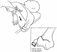
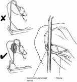
Figure 1.Ulnar nerve compression.Figure 2.Common peroneal nerve compression.
Ulnar nerve injury is more common than brachial plexus injury possibly because of its superficial path along the medial epicondyle of the humerus. The ulnar nerve is particularly vulnerable to compression against the operating table, especially if the forearm is
extended and pronated . Injuries may also occur when the nerve is stretched around the medial epicondyle during extreme flexion of the elbow across the chest There may be certain predisposing factors as suggested by the predominance of this problem in males
(5 : 1), where the cubital tunnel may be narrower, or the ulnar nerve may be unusually mobile. Subclinical lesions may be compounded during surgery as suggested by the finding of abnormal ulnar nerve conduction across the elbow in the unaffected arm .Intra-operative ulnar nerve compression can result in lesions of quite remarkable severity and recovery can be slow and often incomplete. Nevertheless, it seems quite likely that postoperative ulnar nerve palsy can occur without apparent cause and despite accepted methods of positioning and padding
.
Radial nerve
This lesion usually occurs as a result of compression of the nerve between the edge of the operating table and the humerus. It is also the classical `Saturday night palsy'. It may occur when the patient is in the lateral position and the uppermost arm is abducted and suspended from a vertical screen support
.
Sciatic nerve
The sciatic nerve is especially at risk if the patient is thin, the table hard, the operation long and when the opposite buttock is elevated as in the hip pinning position. In the lithotomy position maximal external rotation of the flexed thigh may damage the nerve by stretch .Sciatic nerve palsies have also been reported following coronary artery bypass graft (CABG) surgery, probably as a result of prolonged nerve pressure compounded by the lowered arterial perfusion pressure provided by cardiopulmonary bypass .
Common peroneal nerve Figure 2.
This is the most frequently damaged nerve in the lower limb. It may be compressed against the head of the fibula in the lithotomy position or between the fibula and the operating table, a particular risk associated with the lateral position .
Less common nerve injuries
These include compression of the following nerves: tibial nerve in the popliteal fossa, saphenous nerve between the medial condyle of the tibia and a lithotomy pole, the supraorbital nerve by a tracheal tube or a tight head harness and of the facial nerve , which may be compressed against the ascending ramus of the mandible whilst the anaesthetist holds the jaw forward to maintain an airway.
Prevention of injuries to peripheral nerves in an anaesthetised patient
Prevention of injuries requires an awareness of the potential dangers of the various surgical positions utilised. Careful positioning of every patient on the operating table with proper padding will reduce, but not eliminate, injuries to peripheral nerves. In positions in which strain or pressure on the neurovascular system is possible, the pressure should be alleviated with padding such that the risk of nerve injury is reduced. In preventing brachial plexus lesions, abduction of the arm should preferably be limited . Even with the use of lockable armboards, plexus injury has been reported with the arm abducted to as little as 608 . If 908 abduction is necessary, the elbow should not be fully extended. External rotation of the abducted arm, and especially posterior displacement of the shoulder, should be avoided because this 986
Simplified clinical identification of major peripheral nerve injuries.
(I) Arm
Median Nerve - Numbness over the index finger and Weakness of abduction of the thumb.
Ulnar Nerv ---- Numbness over the little finger, Weakness of abduction and/or adduction of
finger Also, weakness of flexion at the distal interphalangeal joints of the
little and ring fingers the lesion is at the elbow.
Radial Nerve --- Weakness of extension at the distal interphalangeal joint of the thumb,
and of the wrist and finger extensors.
Musculocutaneous Nerve ---- Weakness of flexion of the elbow.
Circumflex Nerve ---- Weakness of abduction of the shoulder.
Brachial Plexus --- Various combinations of lesions within the median, ulnar, radial,
musculocutaneous, and circumflex nerve territories.
(II) Leg
Femoral Nerve ---- Weakness of flexion of the hip. Numbness over the thigh
Obturator Nerve ---- Weakness of adduction of the hip.
Sciatic Nerve --- Weakness of ankle dorsiflexion and plantar flexion. Also, weakness
of knee flexion, if the lesion is proximal. Numbness below the knee.
Common Peroneal Nerve --- Weakness of dorsiflexion of the ankle and toes
Tibial Nerve ---- Weakness of plantarflexion of the ankle and toes.
Stretching can stretch and compress the plexus. Rotation and lateral flexion of the neck towards the opposite side should also be avoided because this increases the tension on the brachial plexus (Fig. 3). Central position of the head is ideal. The severity and permanence of many postoperative lesions of the ulnar nerve make it imperative that every precaution be taken to prevent their occurrence.
Positioning the forearm and hand in pronation places the ulnar nerve at the elbow at risk. Placing the arm in supination and avoiding elbow flexion will free the cubital tunnel . Extremes of elbow flexion should be avoided because this too may cause compression of the ulnar nerve by the medial and arcuate ligaments leading to a neurological deficit. Finally, it seems possible that the ulnar nerve could receive a severe blow from the end of the suspension pole passed up the lateral sleeve of the canvas sheet during patient transfer from operating table to bed (Fig. 4).
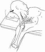
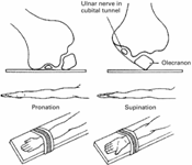
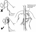
Fig.3 Tension on Fig 4.Ulnar nerve Fig 5. Saphenous nerve
Brachial plexus compression compression.
Since nerve damage to the upper limb can be caused by even minimal compression, the safest position is for the arm to be wrapped in padding down by the patient's side. If this is not possible, maximal padding in the desired position is indicated, with avoidance of other predisposing factors, such as hypothermia and dehydration. This does, however, present a practical problem in having access to an intravenous cannula sited in the hand or arm. We would suggest a free-flowing infusion with a three-way tap sited in the intravenous line to allow rapid administration of intravenous drugs and regular inspection of the cannula site to exclude fluid extravasation. Adequate padding is also required with the patient in the lithotomy position when the saphenous, sciatic and peroneal nerves are vulnerable (Fig. 5).If the patient is in the prone position, the arm should ideally be placed down by the patient's side. If the arms are placed above the patient's head, this can cause a stretch injury to the lower trunks of the brachial plexus. If this is unavoidable, the arm should be placed on an arm board, with the arm abducted to less than 908 with the elbows flexed and the palms facing downwards. Since the commonest injury in the lateral position is to the peroneal nerve, as a result of compression of the lower leg on the table mattress, the nerve must be protected with padding. If the arm is to be suspended, then limitation of arm abduction to 908 is essential. The dependent arm should be anterior to the thorax to avoid compression of the brachial plexus on that side . In the lateral position the head must be properly supported so that the cervical and thoracic portions of the vertebral column are kept in the same horizontal position. Again, this avoids any unnecessary stretch of the brachial plexus. A sound knowledge of nerve anatomy, a cautious injection technique during the block procedure and the judicious use of a nerve stimulator to elicit paraesthesia will also aid in reducing the incidence of nerve injuries. Culpability and medicolegalimplications For nearly a century the literature has contained reports of damage to the peripheral nerves of patients undergoing general anaesthesia. Although these incidents are, potentially, anaesthesia-related injuries, the anaesthetist has not always been to blame, and although purported aetiological factors have been well described, these injuries continue to result in litigation for pain, suffering and economic consequences In some cases there has been a pre-existing lesion or a general increase in susceptibility to damage, and nerve injury occurs despite conventionally accepted methods of positioning or padding. In other cases, the damage has been sustained after the patient has left the operating theatre. it appears that although claims arising from peripheral nerve palsy associated with anaesthesia are rare, when they do occur and the patient suffers permanent and severe neurological deficit, the costs of settling these claims are now well above average anaesthetic claims.
The apportionment of liability between anaesthetist, surgeon and theatre staff is not usually an issue sofar as ulnar nerve palsy is concerned because the anaesthetist is
most often considered to be liable. The uncertain mechanism of ulnar nerve injury implies that the anaesthetist must have done something wrong if the injury occurred in temporal proximity to anaestheticcare . Responsibility for nerve injuries affecting the lower limbs is
difficult to prove. Surgeons, nursing staff and operating department assistants are all involved in manoeuvring the patient into position and pressure or stretching nerve damage can occur at any moment. The apportionment of blame would be a very contentious issue in terms of liability and any subsequent disciplinary procedures. In a claim involving a common peroneal nerve injury, joint responsibility was accepted by surgeon, anaesthetist and theatre staff .
there is need to protect peripheral nerves whilst the patient is anaesthetised and accurately document such care. This will assist in refuting an allegation As with all anaesthetic procedures, meticulous attention to detail is a prerequisite, and an accurately completed anaesthetic chart documenting the peri-operative care is of the utmost importance. While these principles should merely be a reflection of good practice rather than defensive medicine, it is very difficult to defend a claim involving peripheral nerve injury in their absence.
Conclusion
Peripheral nerve lesions are a complication of operative procedures under both general and regional anaesthesia. Although they account for a small proportion of medicolegal claims, they are difficult to defend, beingessentially avoidable. Avoidance involves awareness of the problems associated with operative positions and careful positioning of the patient with appropriate padding. This should form part of standard anaesthetic care. Once damage to nerves has occurred it can take several forms, ranging from a mild, reversible neurapraxia to a permanent sensorimotor deficit. . A carefully completed aesthetic record documenting limb position and appropriate protective measures should be standard for all cases
-------------------------------------------------------------------------------